
Why Is a CBCT Scan Needed Before Dental Implant Surgery?
On this page~10 min read
Key Takeaway
A conventional dental X-ray provides a useful two-dimensional view, but it cannot reliably show the width and three-dimensional shape of the jawbone. A CBCT scan creates detailed 3D images that help the dentist evaluate the implant site, locate important anatomical structures and plan the implant's position, angle and length.
CBCT does involve more radiation than most conventional dental X-rays. Therefore, dentists should only recommend it when the additional information is clinically necessary. The scan area and exposure settings should also be limited to what is needed for treatment planning.
Featured Answer
Do I need a CBCT scan for a dental implant?
A CBCT scan is commonly recommended before dental implant surgery because it shows the height, width and shape of the available jawbone in three dimensions. It also helps the dentist locate nearby nerves, tooth roots, the nasal cavity and the maxillary sinus.
However, imaging should always be selected according to the individual case. The dentist may begin with panoramic or periapical X-rays and recommend CBCT when cross-sectional information is needed for safe and accurate implant planning.
Why Dentists Use CBCT Before Implant Surgery
Patients sometimes wonder why implant treatment requires a CBCT scan when an ordinary dental X-ray costs less. After all, replacing one missing tooth may sound like a relatively simple procedure. The additional imaging can therefore feel like an unnecessary expense.
In reality, a dental implant is placed inside the jawbone. Before surgery, the dentist must understand the shape of the bone and the location of nearby anatomical structures. This information cannot always be obtained from a flat image.
Modern implant treatment also aims to position the implant according to the planned crown. The implant must be surrounded by adequate bone while supporting a restoration that looks natural, functions properly and can be cleaned effectively. A CBCT scan for dental implants gives the clinician three-dimensional information for this planning process.
This does not mean that every dental condition requires CBCT. Routine fillings, uncomplicated extractions and many follow-up examinations can often be assessed with conventional dental X-rays. The scan should answer a specific clinical question that cannot be answered adequately with a lower-dose examination.
Dental X-Rays and CBCT: What Is the Difference?
Periapical and panoramic radiographs are conventional two-dimensional dental images. A periapical X-ray shows a small group of teeth in detail, including their roots and surrounding bone. A panoramic X-ray provides a broader view of the teeth, jaws and nearby structures.
These images remain valuable. Dentists use them to evaluate tooth decay, root infections, periodontal bone loss, impacted teeth and many other conditions. Panoramic and periapical images may also form part of the initial implant assessment.
However, a 2D image compresses three-dimensional anatomy into a flat picture. Structures can overlap, and the image may contain magnification or distortion. Most importantly for implant planning, a conventional radiograph cannot directly show the buccolingual width of the ridge, which is the thickness of bone from the cheek side to the tongue or palate side.
CBCT, or cone-beam computed tomography, uses a cone-shaped X-ray beam while the scanner rotates around the patient. Computer software reconstructs the collected data into a three-dimensional image. The dentist can inspect the area from different directions and examine cross-sectional slices of the jaw.
| Imaging method | Type of image | Information provided | Common implant-related use |
|---|---|---|---|
| Periapical X-ray | 2D | Detailed view of several teeth, roots and nearby bone | Initial assessment and implant follow-up |
| Panoramic X-ray | 2D | Broad view of both jaws and major structures | General screening and preliminary planning |
| CBCT scan | 3D | Bone dimensions and spatial relationships between anatomical structures | Cross-sectional implant-site assessment and surgical planning |
The American Academy of Oral and Maxillofacial Radiology states that panoramic imaging may be used for the initial assessment. However, it recommends cross-sectional imaging for preoperative implant-site evaluation, with CBCT as the current method of choice when that information is required. Source: AAOMR position statement
Implant Placement Requires Three-Dimensional Planning
A dental implant is an artificial tooth root placed into the alveolar bone. It must have enough surrounding bone for appropriate stability and long-term support. Therefore, the clinician needs to evaluate more than the visible gap between adjacent teeth.
The jawbone may appear tall enough on a panoramic X-ray while being too narrow in another dimension. Bone loss after extraction can also produce an irregular ridge. A concavity on the inner or outer surface may not be obvious on a two-dimensional image.
CBCT allows the dentist to study:
- The height and width of the available bone
- The shape and inclination of the residual ridge
- The relationship between the implant site and adjacent roots
- The location of the mandibular canal and mental foramen
- The position of the maxillary sinus and nasal floor
- Possible impacted teeth, lesions or other unexpected findings
These details influence the proposed implant diameter, length, angle and position. They may also show whether bone grafting, sinus augmentation or another site-development procedure should be considered.
CBCT measurements should still be interpreted together with a clinical examination, medical history and restorative plan. A scan alone does not determine whether someone is suitable for implant treatment.
Protecting Nerves, Sinuses and Adjacent Teeth
The jaws contain several structures that must be considered during surgery. In the lower jaw, the inferior alveolar nerve travels through a canal inside the bone. Injury to this area may cause altered sensation or numbness affecting the lower lip and chin.
In the upper posterior jaw, the roots of the teeth and proposed implant sites may be close to the maxillary sinus. Bone height can become limited after tooth loss because the ridge resorbs and the sinus may extend downward. The nasal floor also matters when implants are planned in the anterior upper jaw.
Adjacent natural teeth create another limitation. The implant needs sufficient separation from their roots and from other implants. Poor positioning may affect the bone, soft tissue, restoration and the patient's ability to clean the area.
Because CBCT displays these relationships in three dimensions, it helps the dentist plan a safer surgical pathway. In suitable cases, the data can also support virtual implant planning and the production of a surgical guide. CBCT cannot remove every surgical risk. Image quality, patient movement, metal artifacts and diagnostic interpretation all affect its usefulness. Safe treatment still depends on professional training, clinical judgment and careful surgery.
Can CBCT Measure Bone Quality?
CBCT is very useful for assessing bone dimensions and morphology. It can show cortical boundaries, ridge defects and the relationship between the implant site and nearby anatomical structures.
However, patients may hear that CBCT provides an exact measurement of bone density. This requires clarification. Gray values from dental CBCT are not standardized in the same way as Hounsfield units from conventional medical CT. They can vary among scanners and imaging settings.
Therefore, CBCT should not be presented as a perfect or universal test of implant-site bone quality. The dentist may form an initial impression from the scan, but final bone characteristics and implant stability are also assessed clinically during surgery.
This correction is important because a detailed-looking image does not make every measurement equally reliable. The strongest role of CBCT in implant planning is its ability to show three-dimensional anatomy and available bone dimensions.
Is Dental CBCT Radiation Safe?
CBCT uses ionizing radiation, so describing it as completely risk-free would be inaccurate. A dental CBCT examination generally exposes the patient to more radiation than a conventional periapical or panoramic dental X-ray.
At the same time, dental CBCT usually delivers less radiation than conventional medical CT examinations of larger anatomical areas. The actual dose can vary considerably according to the scanner, field of view, image resolution, exposure settings and patient size. For this reason, one fixed dose cannot accurately represent every dental CBCT examination.
The correct approach is justification and optimization:
- Order the scan only when it is expected to provide clinically useful information.
- Use the smallest field of view that covers the area of interest.
- Select exposure settings appropriate for the patient and diagnostic task.
- Review previous images to avoid unnecessary duplicate examinations.
- Use special caution with children and younger patients.
The FDA explains that CBCT provides three-dimensional information that may assist diagnosis and treatment planning. It also notes that CBCT usually involves more radiation than conventional dental X-rays and should only be performed when necessary. Source: U.S. Food and Drug Administration
For a properly selected patient, the expected diagnostic and treatment-planning benefits generally outweigh the small radiation risk. However, the decision must be individualized rather than based on a routine one-scan-for-everyone policy.
When Is an Ordinary Dental X-Ray Enough?
Conventional dental radiographs have not been replaced by CBCT. They remain the first choice for many common dental situations because they provide useful information with lower radiation exposure and lower cost.
A periapical image may be appropriate for examining a tooth after implant placement, checking bone levels during follow-up or evaluating a localized dental problem. A panoramic image may offer a helpful initial overview of the jaws and existing teeth.
CBCT becomes more valuable when the dentist needs information that a flat image cannot provide. Examples include evaluating the width of an implant site, locating a nerve canal, studying the maxillary sinus or planning grafting and guided surgery.
The choice should therefore be based on the clinical question rather than price or technology alone. A more expensive image is not automatically better, and a less expensive image is not automatically sufficient.
What Happens During a CBCT Scan?
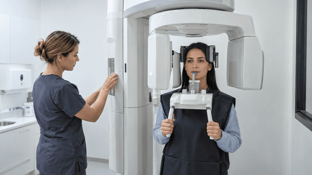
A dental CBCT examination is usually brief and noninvasive. Depending on the machine, the patient may stand or sit while the scanner rotates around the head. The patient must remain still to reduce movement and preserve image quality.
Before the scan, staff may ask the patient to remove glasses, jewelry, removable dental appliances or other metal objects around the head and neck. Metal can create artifacts that make parts of the image more difficult to interpret.
The scan itself usually takes less than a minute, although positioning and preparation may take longer. The computer then reconstructs the information into a 3D dataset for evaluation.
Afterward, the clinician reviews the entire captured volume, not only the missing-tooth area. This is important because the scan may reveal findings elsewhere in the jaws, sinuses or surrounding structures that require attention.
Why Accurate Imaging Can Improve Implant Planning
Better imaging does not guarantee implant success, but it reduces uncertainty during treatment planning. CBCT can help the clinician select a position that respects anatomical limits while supporting the planned crown.
It can also reveal conditions that require a change in treatment. For example, the scan may show inadequate ridge width, sinus involvement, an unexpected lesion or an unfavorable relationship with an adjacent root. The dentist can then discuss grafting, an alternative implant position or a different restorative option before surgery begins.
Accurate planning may also help limit unnecessary surgical exploration. When the anatomy and restorative goals are understood in advance, the procedure can be planned more deliberately.
Implant success still depends on many other factors, including periodontal health, plaque control, smoking, diabetes management, bite forces, implant stability and follow-up care. CBCT is an important planning tool, but it is only one part of a complete assessment.
Frequently Asked Questions
Can dental implants be done without a CT scan?
Implants have historically been placed using clinical assessment and conventional radiographs. However, a 2D image cannot show the complete width and shape of the implant site. Current professional guidance supports cross-sectional imaging when it is needed for accurate preoperative assessment, with CBCT commonly used for this purpose.
How much radiation is in a dental CBCT scan?
There is no single dose for every CBCT scan. Exposure varies with the machine, field of view, resolution and patient settings. It is generally higher than conventional dental radiography but lower than many medical CT examinations. The smallest clinically appropriate scan area should be used.
Conclusion
A panoramic or periapical dental X-ray remains useful for many examinations, but it only presents anatomy in two dimensions. Implant placement occurs within a three-dimensional jaw that contains nerves, sinuses, tooth roots and bone surfaces that may overlap on an ordinary image.
A CBCT scan for dental implants helps the clinician assess bone dimensions, understand the surrounding anatomy and plan the implant's position more accurately. It should not be ordered without a clinical reason, and exposure should be kept as low as reasonably achievable.
When your dentist recommends CBCT before implant surgery, ask what information the scan is expected to provide and whether a limited field of view can be used. A clear explanation should connect the imaging examination directly to your individual treatment plan.
Planning Dental Implants and Need a CBCT Assessment?
Send Huangshan Dental your dental X-rays or CBCT images for an initial review. Our team can help assess your available bone, nearby anatomical structures and possible implant treatment options before you arrange your visit.