DEXIS ,Designed to increase practice efficiency, the DEXIS OP 3D imaging system combines excellent image quality with customizable features to meet the practice’s diagnostic needs, , ,Greg Gillespie, DDS
,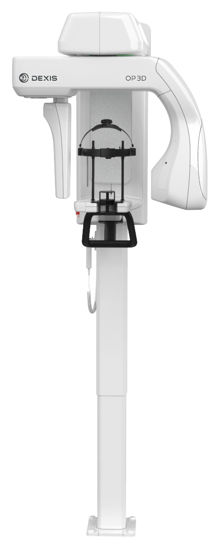 A picture is worth a thousand words. In dentistry, that image should provide the data to eliminate guesswork and deliver predictability. “If an imaging unit can’t capture a great image, I don’t care about its bells and whistles—it’s not going to be worth anything,” said Greg Gillespie, DDS, who uses the DEXIS ORTHOPANTOMOGRAPHTM OP 3DTM imaging system in his Vancouver, WA, practice., ,While the DEXIS OP 3D is certainly comprehensive, it’s specifically designed for excellent image quality and clinical efficiency through features such as intuitive patient positioning and a graphical user interface., ,When it comes to diagnosis, the cone beam 3D imaging unit, which also offers panoramic and cephalometric imaging, features a variety of image resolutions and FOV sizes that meet the diagnostic needs of general dental practitioners, surgeons, and specialists. Metal artifact reduction (MAR) is activated within all FOV sizes to provide a high level of image quality in all cases, including endodontics and implant placement., ,“The image quality of the DEXIS OP 3D is fantastic,” said Dr. Gillespie. “It makes my diagnosis quick and more precise because I can see the image very well in many different positions. Once you have this unit, you realize, ‘Oh my goodness, I can’t diagnose without it.’”, ,For Gillespie Dentistry, the DEXIS OP 3D has become a key diagnostic tool for virtually any clinical scenario. “One of our best diagnostic tools in hygiene is that we have patients take a full scan every 5 years, including imaging of all the root canals that have been previously treated,” he shared. “I can’t tell you how many times we’ve found a root canal with an infection that we couldn’t see with a 2D image, but it shows up very clearly in a 3D image.”, ,When planning for wisdom teeth extraction, the DEXIS OP 3D allows Dr. Gillespie to see where the mandibular nerve lies in relation to the tooth. By rotating an image 3-dimensionally, he can visualize how intimately the tooth’s root tips are involved with the nerve, which creates a much safer approach toward treatment., ,Lastly, during implant planning, the DEXIS OP 3D allows him to very clearly visualize bone quality and quantity. On the software side, DEXIS’ DTX Studio allows Dr. Gillespie to overlay this 3D imaging data with the patient’s digital scan and then view all of the images together in one place., ,“This makes your planning incredibly accurate so that when you place the implants, they come out exactly as you anticipated,” Dr. Gillespie said. “Essentially, the questions go down, and your predictability goes up.”, ,Dr. Gillespie received his dental degree from the University of Washington School of Dentistry in Seattle, WA. He maintains a full-time dental practice in Vancouver, WA, which focuses on general dentistry with an emphasis on implant and cosmetic dentistry. His vision of comprehensive dentistry involves effective treatment planning and use of the best dental materials available. Dr. Gillespie lectures across the country and participates in ongoing evaluations of the latest materials and techniques in dentistry.
