My in-office CBCT imaging unit has been the foundation for my complete dental examination since 2009. With my purchase of GALILEOS from Dentsply Sirona, I became a clairvoyant (one who sees clearly), routinely deciphering problems thanks to the 3D difference its imaging provides., ,The following case report illustrates how 3D imaging elevates my diagnostic and treatment planning ability, thereby leading to improved clinical decisions., ,A 27-year-old female in good health presented for a consultation regarding 2 areas of concern. She wanted options to replace an over-retained primary upper right canine, and asked if she was a candidate for Invisalign. I quickly determined that she could improve her occlusion and smile with Invisalign. Replacing the primary canine was a little trickier, as the permanent canine was vertically impacted and multiple dentists recommended the removal of the impacted tooth prior to considering implant placement., ,Diagnosis and Treatment Plan, ,The initial diagnostic information was gathered by clinical examination, periodontal evaluation, and exposure to Schick 33 radiography from Dentsply Sirona. I followed this with an enhanced 3D analysis via GALILEOS, which gave me the necessary information to make a proper diagnosis. Reviewing the 3D data put her at ease, because we evaluated the maxillofacial anatomy on a large monitor and intelligently discussed all options available for a fixed tooth replacement. Initial evaluation using a virtual implant provided certainty that an implant could be installed without disturbing the impacted canine. The 3D imaging revealed clear evidence that an invasive oral surgery could be avoided, and the goal of replacing the primary canine was possible via computer-assisted guided implantology, which became the treatment choice for this patient., ,Additional information was gathered with CERECGALILEOS integration, where the optical digital scan and the 3D radiographic scan were merged. Full control of all aspects of implant dentistry, both surgically and restoratively, was achieved. My patient could now undergo less invasive, safe, and precise implant placement via flapless guided surgery., ,The digital workflow began with a GALILEOS CBCT scan, followed by an optical scan of her dental anatomy with Omnicam from Dentsply Sirona. These 2 scans were merged and a restoratively driven virtual implant was planned using GALILEOS implant software. Care was taken to avoid penetrating the impacted canine. A SICAT optiguide was manufactured to guide the osteotomy and implant installation during the flapless guided implant surgery., ,Implant Placement, ,The actual surgical visit took less time than it takes to excavate and restore a 2-surface resin restoration. The NobelActive implant from Nobel Biocare was placed according to plan and was immediately provisionalized with a screw-retained Telio hybrid abutment crown that was fabricated digitally in CEREC and milled out in my inLab MC XL milling machine. The patient remarked how easy the implant surgery went and was thrilled with our treatment. An Osstell Resonance Frequency Analysis reading of 70 was recorded, which means superior initial stability was achieved, verifying objectively that this implant could be loaded. GALILEOS imaging validated my clinical decision and led to a minimally invasive guided implant treatment., ,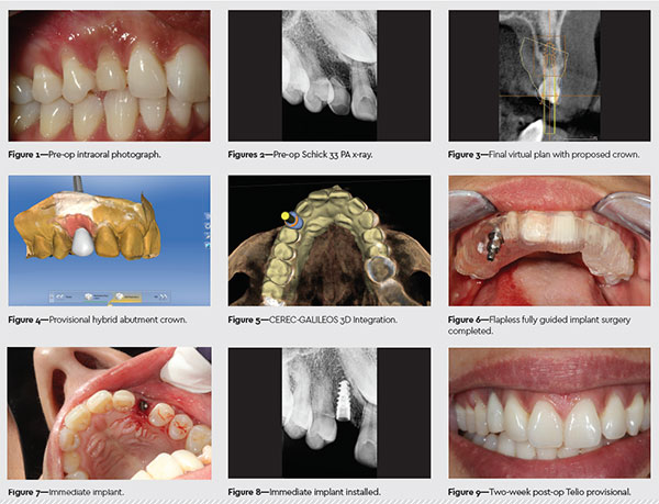 ,
,
/
/
Guided Implantology to Replace Over-Retained Primary without Removal of Impacted Canine
