Body
Presentation
Population
- Patients with previous extensive restorations (e.g., amalgam or resin restorations, crowns), trauma or recurrent caries with pulp exposure
- Medically-compromised patients
Signs
- Swelling (sometimes)
- Sinus tract (sometimes)
- Large periapical lesions (sometimes)
Symptoms
- Pain severity may vary
- Continuous flow of pus or serous-like fluid
- Bad taste in mouth (drainage from sinus tract)
Investigation
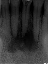 Fig. 1: Pre-treatment radiograph: large periapical radiolucency associated with two necrotic teeth.
- Thoroughly assess the patient’s medical history: diabetes mellitus, bleeding disorders, history of radiation therapy and trauma.
- Perform a complete extraoral and intraoral examination:
- Examine for sinus tract inside or outside the oral cavity. If present, trace radiographically.
- Examine for swelling and periodontal pocketing.
- Examine teeth for caries, broken down restorations, crowns with open margins or recurrent caries.
- Perform pulp tests (hot, cold and electric) on the tooth in question and surrounding teeth to ensure they are not contributing to the problem.
- Perform a radiographic examination:
- periapical radiographs to check for periapical (PA) pathology and periodontal problems (Fig. 1)
- bitewing radiographs to check for dental caries
- Perform a radiographic examination to investigate:
- recurrent caries
- pulp exposures
- widened periodontal ligaments
- external and internal resorption
- calcifications
- length accuracy, perforations, strip perforations, or possible additional canals after the initiation of treatment
- pathology in the area around the teeth or in the maxillary sinus
Diagnosis
Based on the clinical and radiographic examinations and the patient’s medical history, a diagnosis of necrotic tooth with unstoppable drainage is determined.
Treatment
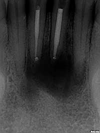 Fig. 2: Post-treatment radiograph: treatment involved both non-surgical and surgical endodontic procedures to resolve the unstoppable drainage.
If the patient presents with a fluctuant swelling, consider doing an incision and draining prior to initiating treatment. Then begin instrumentation.
- Perform a more thorough cleaning and shaping of the canal spaces to ensure that all necrotic materials have been removed.
- Verify length determination (apex locator and radiographs) to ensure that over-instrumentation did not occur. Special care should be taken near the maxillary sinus, since over-instrumentation can lead to persistent drainage.
- If a strip perforation or perforation is noted, repair immediately with MTA or equivalent material. If unable to perform this procedure, refer the patient to an endodontist.
- Irrigate with NaOCl and leave in the canals and chamber for 10–15 minutes. Dry and place Ca(OH)2 in the canals and close, if drainage stops.
- Use negative pressure irrigation, if available.
- If all else fails, leave the tooth open, reappoint the next day, lightly instrument, irrigate and dry, and close the canal.
- In cases where there is a large PA radiolucency associated with a necrotic tooth and the drainage continues, both conservative and surgical endodontic treatments may be required (Fig. 2). Refer to an endodontist if uncomfortable dealing with this situation.
THE AUTHOR
Suggested Resources
- Imura N, Zuolo ML. Factors associated with endodontic flare-ups: a prospective study. Int Endod J. 1995;28(5):261-5.
- Morse DR, Koren LZ, Esposito JV, Goldberg JM, Belott RM, Sinai IH, et al. Asymptomatic teeth with necrotic pulps and associated periapical radiolucencies: relationship of flare-ups to endodontic instrumentation, antibiotic usage and stress in three separate practices at three different time periods. Int J Psychosom. 1986;33(1):5-87.
- Walton R, Fouad A. Endodontic interappointment flare-ups: a prospective study of incidence and related factors. J Endod. 1992;18(4):172-7.
- Harrington GW, Natkin E. Midtreatment flare-ups. Dent Clin North Am. 1992;36(2):409-23.
- Harrison JW, Gaumgartner JC, Svec TA. Incidence of pain associated with clinical factors during and after root canal therapy. Part 1. Interappointment pain. J Endod. 1983;9:384.
- Fabricius L, Dahlén G, Sundqvist G, Happonen RP, Möller AJ. Influence of residual bacteria on periapical tissue healing after chemomechanical treatment and root filling of experimentally infected monkey teeth. Eur J Oral Sci. 2006;11(4):278-85.
- Seltzer S, Bender IB, Ziontz M. The dynamics of pulp inflammation: correlations between diagnostic data and actual histologic findings in the pulp. Oral Surg Oral Med Oral Pathol. 1963;16:846-71.
- Tsesis I, Faivishevsky V, Fuss Z, Zukerman O. Flare-ups after endodontic treatment: a meta-analysis of literature. J Endod. 2008;34(10):1177-81.
- Torabinejad M, Kettering JD, McGraw JC, Cummings RR, Dwyer TG, Tobias TS. Factors associated with endodontic interappointment emergencies of teeth with necrotic pulps. J Endod. 1988;14(5):261-6.
Request reprint permission
