
The following clinical case report presents the successful treatment of bilateral sites using both immediate and conventional implant placement with the Straumann Fast Molar Solution. The Straumann Anatomic Healing Abutment XC facilitated optimal soft-tissue conditioning and enhanced patient comfort, while its scannable head introduced a new level of time efficiency by eliminating the need for abutment removal.
Initial situation
A 38-year-old female patient presented to the dental office reporting social discomfort when smiling, owing to the absence of both maxillary second premolars (Figs. 1a– c, Figs. 2a–c & 3). Clinical examination revealed that tooth #16 had an intra-radicular post and had been prepared for a crown, but the crown was missing, and a cavity was noted on the distal surface of tooth #14. Overall, the patient exhibited a healthy periodontal condition. She was a non-smoker and did not present with any comorbidities or systemic health issues. CBCT imaging showed adequate bone quality and quantity at both sites and no signs of acute infection. Additionally, a residual root was identified at site #15 beneath the already healed gingiva.

Figs. 2a–c: Intra-oral images of the initial situation.
Treatment planning
The patient’s maxillary and mandibular arches were scanned using the Straumann SIRIOS system. The scans, along with the DICOM files from the CBCT scan, were sent via Straumann AXS to Smile in a Box (Straumann) for treatment planning, surgical guide design and 3D printing of the model and surgical guides (Figs. 4, 5a–f & Figs. 6a–d).
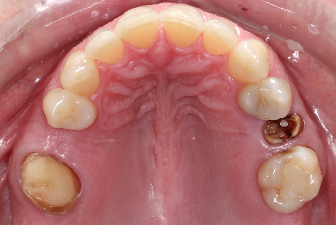
Fig. 3: Occlusal view of the initial situation.
After evaluation and validation of the plan, it was decided for site #15—where a residual root was present and the bone density was generally soft—that a Straumann BLX (Roxolid, SLActive, regular base) 4.5 × 8.0 mm implant would be placed, along with an M shape Straumann Anatomic Healing Abutment XC (regular base/wide base; gingival height: 1.5 mm). For site #25, extraction of the remaining root was planned and would be followed by the placement of a Straumann BLC (Roxolid, SLActive, regular base) 3.75 × 8.00 mm implant. This narrower site had a visible lamina dura, favourable for achieving primary stability. An M shape Straumann Anatomic Healing Abutment XC (regular base/wide base; gingival height: 1.5 mm) was also selected for this site to support proper soft-tissue emergence during the 60- to 90-day healing period. Owing to the expected post-extraction gaps between the implants and buccal bone walls, cerabone plus (botiss biomaterials) was planned to be used for grafting.
After the healing and gingival maturation phases, the patient would return for intra-oral scanning directly on the healing abutments, eliminating the need for abutment removal thanks to their scannable head design. Definitive restorations were planned as full-contour monolithic zirconia crowns cemented on to Straumann Variobase abutments.


