INTRODUCTION
Sjögren’s Syndrome (SS) is a systemic autoimmune disease affecting the exocrine glands. It is characterized by intense lymphocytic infiltration that progressively destroys mainly salivary and lacrimal glands.1,2 SS is subdivided into 2 categories: primary, which includes dry eyes and hyposalivation, and secondary, which occurs in conjunction with other connective-tissue diseases (eg, arthritis and lupus erythematosus, among others).3 The etiology is unknown. It is suggested that an interaction between environmental factors (eg, viruses, stress, hormones) and patient genetics can lead to inflammatory responses against epithelial tissues.4,5
SS is the second most common connective tissue disease, affecting up to 3.1 million Americans, or approximately 1 in 70 people, according to the National Arthritis Data Workgroup. This number refers to those presenting with primary SS. It can be doubled if including patients presenting with secondary SS.6,7 It is more commonly found in women than in men (9:1) and is most often diagnosed during the fourth and fifth decades of life.5,8
Oral symptoms include dry mouth; cracked lips; angular cheilitis; mucosal sores; tongue depapillation; swelling of the salivary glands; oral infections, such as candidiasis; and difficulty in mastication, swallowing, and speech. Hyposalivation and changes in saliva composition can cause a burning sensation and an intolerance to the use of removable prostheses.9 Moreover, patients with xerostomia have a high incidence of caries, resulting in the extraction of teeth during their lifetimes.10 This oral agony impacts patients psychologically and emotionally, affecting their social interactions and quality of life.11

Early interdisciplinary intervention is recommended to provide long-term solutions for improving function, speech, aesthetics, and self-esteem.
Oral rehabilitation with implants is a possible solution since SS does not affect bone healing and osseointegration.2,5,12 Binon and Fowler13 presented a case report with mandibular-osseointegrated implants and a fixed prosthesis that remained functional and stable after 13 years of followup. Due to the difficulties SS patients have wearing removable prostheses, a fixed transitional prosthesis during the surgical stage is key. Drew et al14 proposed the stage approach, in which hopeless teeth are kept in strategic positions as abutments to support an interim fixed prosthesis during the surgical stage, making the transition from a terminal dentition to a full-arch reconstruction easier. Some of the advantages of this technique include increased patient comfort, soft-tissue management, protection of grafted areas, alveolar bone preservation, and surgical templates for the position of future implants.
Furthermore, the severity of caries makes the extraction of teeth a very common procedure affecting the alveolar ridge morphology. In general, the buccal vertical bone resorbed 2.2 mm after extraction, making the ridge augmentation procedure required before implant placement in most cases. Another option to avoid extraction in strategic sites is root preservation. Its requirements include prophylactic root canal therapy and removal of coronal tooth structure, leaving the root face below the crestal bone. This technique preserves the ridge form on pontic sites between teeth or implants, supports the soft tissue, and prevents epithelial down growth.15 The aim of the present case series is to show a simple way to restore these complex cases. Case 1 shows the sequence from a terminal dentition to full-mouth, implant-supported prostheses using the stage approach and the root banking technique. Case 2 shows the longevity of an SS patient after 15 years of followups using the staged approach.
CASE REPORTS
Case 1
A 59-year-old Caucasian female presented to the department of periodontics, Rutgers School of Dental Medicine, with the chief complaint of “All my teeth are breaking.” She was diagnosed with SS and rheumatoid arthritis in 2015. The patient was dissatisfied with her smile and came to the school to find a permanent solution. Intraoral examination revealed a partially edentulous maxilla and mandible, dry mouth, and subgingival carious lesions in the majority of her teeth with a decrease in the occlusal vertical dimension (OVD) (Figure 1). Radiographic examination revealed sinus pneumatization, recurrent and subgingival caries, and defective endodontic lesions (Figures 2 and 3). On the first visit, oral health was addressed by prescribing a high-fluoride toothpaste (PreviDent 5000 [Colgate Oral Pharmaceuticals]) to be used 3 times per day. A comprehensive evaluation of her case with a multidisciplinary team, including the prosthodontics and periodontics departments, determined that a full-mouth implant rehabilitation would be the best option for her. The patient was motivated and agreed to proceed with the treatment.


Treatment was planned to have multiple phases. The first phase involved the abutment selection to hold the interim fixed prostheses. In the maxilla, the upper right canine, central incisors, and first upper left premolar were selected, and in the mandible, the lateral incisors, lower left first molar, and lower right second premolar were selected. These teeth presented a strategic position in the arches without periodontal involvement or mobility. Preliminary casts were articulated in centric relation, and diagnostic waxing was made with OVD increased, improving aesthetics and function.
The teeth were prepared, and hopeless teeth were decoronated (their pulp chambers were calcified). Subgingival carious lesions were excavated and restored with glass ionomer material. Then polymethyl methacrylate (PMMA) shells with metal reinforcements were relined with PMMA (ALIKE [GC America]) (Figures 4 to 6).


The second step involved the surgical phase. Decoronated teeth were extracted and grafted with allograft (RegenerOss [ZimVie]) and resorbable membrane (Geistlich Bio-Gide [Geistlich Pharma AG]) to maintain the bony architecture for future implant placement (Figure 7). Four months later, CBCT was taken with guide stents. Implant planning was made with Romexis planning software (Planmeca Group), and tooth-borne surgical guides were fabricated (Figure 8). Five implants (ZimVie) were placed in each arch: In the maxilla, 4/3- × 10-mm upper right first premolar, 3.25- × 13-mm and 3.25- × 11.5-mm lateral incisors, 3.25- × 11.5-mm upper left canine, and 5/4- × 10-mm second premolar; and in the mandible, 4/3- × 8.5-mm implants in the lower left second premolar and canine, 3.25- × 10-mm between central incisors, and 4/3- × 10-mm in the lower right first premolar and first molar sites (Figures 9 and 10). After surgery, interim fixed prostheses were adjusted, and pressure was released against the tissues. Implant sites were uncovered after 4 months. Then digital scans were made (TRIOS [3Shape]) with scan bodies (BellaTek Encode Impression System [ZimVie]) (Figures 11 and 12). Scans were imported into a CAD/CAM software program (3Shape Dental System [3Shape]) to fabricate a new set of interim prostheses.


The third phase involved prophylactic root canal therapy in the upper right canine, central incisor, and lower lateral incisors. Root canal therapies were performed, the coronal gutta-percha was removed, and tapered preparation of the coronal canal space was made to facilitate the Geristore (DenMat) placement 2 mm below the crestal bone. A minimum of 5 mm of gutta-percha was maintained in the apical portion of the canal space to ensure an adequate apical seal. Geristore was placed in the tapered wall preparation with a small tip engaged at the level of the gutta-percha (Figure 13). The remaining teeth were decoronated, and full-arch, screw-retained prostheses were inserted, making the transition from tooth-borne interim prostheses to implant-supported interim prostheses. The upper left central incisor, second premolar, lower left first molar, and lower right second premolar were extracted and grafted with allograft (RegenerOss [ZimVie]).


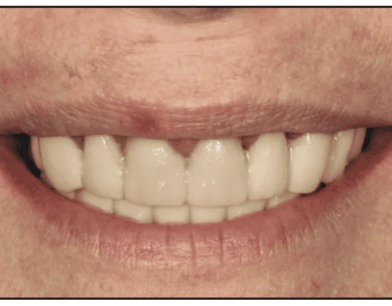
The fourth step involved the restorative phase, in which maxillary and mandibular master casts were generated and verified. Gold-shaded titanium custom abutments (Atlantis [Dentsply Sirona]) were inserted in the maxilla, and multi-unit abutments were placed in the mandible. PMMA prototypes were inserted and adjusted. The patient stayed with the prototypes for 2 weeks to evaluate oral hygiene, phonetics, aesthetics, and function. All the corrections were made to them. After 2 weeks, prototypes were re-scanned in the laboratory. Definitive monolithic zirconia prostheses were delivered (Figures 14 and 15). An occlusal device was inserted with 4 months of recall appointments. A treatment plan sequence was summarized in the schematic view using Dental_Flash (Attachments International) (Figures 16 and 17).
Case 2
A 62-year-old white female presented to the periodontics department in August 2006. Her chief complaint was that she was “tired of fillings and root canal treatments.” Previous dental treatment included several extractions, root canals, implants, and a removable partial prosthesis that she did not tolerate. She stated she had been through several reconstructions that had failed due to caries on abutment teeth. She was diagnosed with SS and Lichen Planus many years ago. Intraoral examination revealed generalized gingival inflammation with bleeding on probing, crowns and bridges in the maxilla, subgingival caries, and defective restorations in the mandible (Figure 18). Radiographic examination revealed defective restorative materials, RCTs, carious lesions, and implants on the molars and premolar areas (Figures 19 and 20). After evaluation of all her factors and patient expectations, the option of implant-supported, fixed prostheses was offered to the patient. The stage approach was crucial in her case because she had poor tolerance of a removable prosthesis in the past. A schematic sequence using Dental_Flash briefly summarized her phases using the staging approach (Figures 21 and 22). The patient has been stable for the past fifteen years with 4-month recall appointments (Figure 23).


