
A cavity, also called dental caries, is the result of tooth decay. It develops when plaque builds up and produces acid that erodes enamel. If not treated, it can cause damage to deeper layers of the tooth, leading to infection or tooth loss.
When dentists suspect a cavity, they often confirm it using X-rays. But what does a cavity look like on an X-ray? This article offers a detailed explanation to help you understand the signs of decay on dental radiographs. It will also touch on related conditions such as sensitive teeth and gum disease.
Why Dentists Use X-Rays to Detect Cavities
X-rays are essential in dentistry. They allow professionals to detect problems that aren’t visible to the naked eye. Cavities can form between teeth, under fillings, or beneath the surface of enamel. X-rays, especially bitewing radiographs, are key tools for spotting these hidden issues.
The American Dental Association (ADA) recommends regular X-rays to catch cavities in their early stages.
Types of Dental X-Rays and Their Role
There are several types of dental X-rays, each with a unique function:
Bitewing X-rays: Show upper and lower back teeth and are commonly used to detect cavities between teeth.
Periapical X-rays: Capture the entire tooth from crown to root and are useful for assessing abscesses and severe decay.
Panoramic X-rays: Show a broad view of the jaws, used less frequently for cavity detection but helpful in evaluating overall oral health.
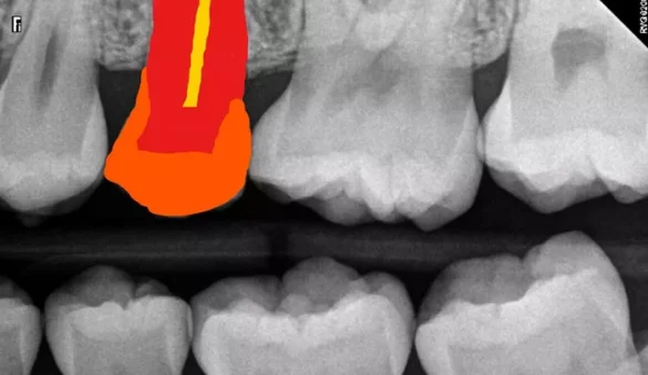
What a Cavity Looks Like on an X-Ray
On an X-ray, healthy tooth enamel appears as a light or white area because it is dense and resists radiation. A cavity shows up as a dark or radiolucent spot due to demineralization and loss of density.
Depending on the stage and location of the cavity, the image might show:
Dark lines or shadows between teeth — typical of early decay in the enamel.
Larger dark areas penetrating the dentin — indicate deeper decay.
Dark zones near fillings — may suggest recurrent decay under or around existing restorations.
X-rays help determine the extent of the cavity, guiding treatment options like fillings, crowns, or root canals.
How Deep Cavities Affect Oral Health
If a cavity reaches the inner layers of the tooth, it can lead to inflammation of the pulp and increased sensitivity. Many patients with sensitive teeth are surprised to find that the underlying cause is a hidden cavity revealed on X-ray.
Untreated cavities may also contribute to gum disease. When decay spreads below the gumline or affects adjacent gum tissues, it can lead to irritation, bleeding, and periodontal issues.
Stages of Tooth Decay on X-Ray
Tooth decay progresses through several stages, each with distinct X-ray features:
Enamel Decay: Small dark spot near the surface. Treatment may include fluoride or sealants.
Dentin Involvement: Cavity extends deeper, requiring a dental filling.
Pulp Infection: Involves the nerve chamber, possibly needing a root canal.
Abscess Formation: Visible as a dark halo around the root, indicating infection spread.
Importance of Regular X-Rays for Prevention
Regular dental check-ups combined with routine X-rays can catch cavities early. This proactive approach minimizes invasive treatments and preserves your natural teeth.
Patients with a history of sensitive teeth or gum disease may benefit from more frequent X-ray monitoring. Early detection reduces the risk of tooth loss, pain, and costly procedures.
Other Dental Conditions Seen on X-Rays
In addition to cavities, X-rays can reveal:
- Bone loss associated with gum disease
- Impacted wisdom teeth or other eruptive issues
- Infections or abscesses around roots
- Cysts or tumors in the jaw
This makes X-rays a valuable tool in comprehensive dental care planning.
How Sensitive Teeth and Gum Disease Interact with Cavities
Sensitive teeth can result from many causes, including worn enamel, exposed roots, and tooth decay. When X-rays reveal cavities in patients with sensitivity, targeted treatment can relieve discomfort and prevent further damage.
Gum disease often coexists with cavities. As gum tissue recedes, tooth roots become more exposed and vulnerable to decay. X-rays allow the dentist to assess both the bone and the teeth to create a comprehensive treatment strategy.
Treatment Options Based on X-Ray Findings
Treatment depends on the cavity’s depth and location as seen on the X-ray:
Small cavities: Remineralization or small composite filling.
Moderate decay: Composite or amalgam filling.
Deep cavities: May require a root canal and crown.
Extensive decay: Tooth extraction and replacement options like implants.
Identifying the correct treatment relies heavily on interpreting X-ray images accurately.
Can You Spot a Cavity Yourself on an X-Ray?
While it’s tempting to try, interpreting dental X-rays requires training. A cavity may be subtle and hard to identify without experience. If you have symptoms like tooth sensitivity, pain, or visible holes, see your dentist and let them review your X-rays.
Still, patients who understand what cavities look like on an X-ray can ask better questions and make more informed decisions about their care.
Frequently Asked Questions
Can a cavity be missed on an X-ray?
Yes. Small or early-stage cavities may not always appear clearly. Dentists also rely on visual exams and probing with dental instruments.
How often should dental X-rays be taken?
It depends on your age, oral health, and history of cavities. Most adults get bitewing X-rays every 1-2 years, more often if there is gum disease or frequent decay.
Are dental X-rays safe?
Yes. Modern digital X-rays use very low radiation doses. Dentists take precautions, especially with children and pregnant women.
Conclusion
Understanding what a cavity looks like on an X-ray empowers you to take charge of your oral health. If you deal with sensitive teeth or a history of gum disease, early detection is even more important.
By maintaining regular visits and being proactive, you can avoid serious dental issues and preserve your smile for years to come. X-rays are a key part of this preventive strategy.