
For me, practicing dentistry has always been exciting and fun, yet has also presented challenges, especially when I’ve thought that I might not be able to do the best job. My patients are the most important asset in my practice, and it is essential to provide them with the best possible care. Modern technology allows me to do exactly that: Achieve the optimal outcome predictably and safely, making the patient experience exceptional.
The technology that has had the greatest influence on the patient experience is cone-beam imaging. I was first exposed to this technology in 2014, and soon after, I successfully implemented implant placement in my practice. Recently, we added Dentsply Sirona’s newest 3D/2D unit, Axeos. My first impression was that it has a much smaller footprint than other systems we’ve used. Second, Axeos is a superbly designed piece of art with built-in LED lights—patients always comment on the changing colors it emits. Third, and most important for us clinicians, is the quality, clarity, and resolution of the images, which are of a diagnostic quality that I have never experienced.
Planning with the Patient
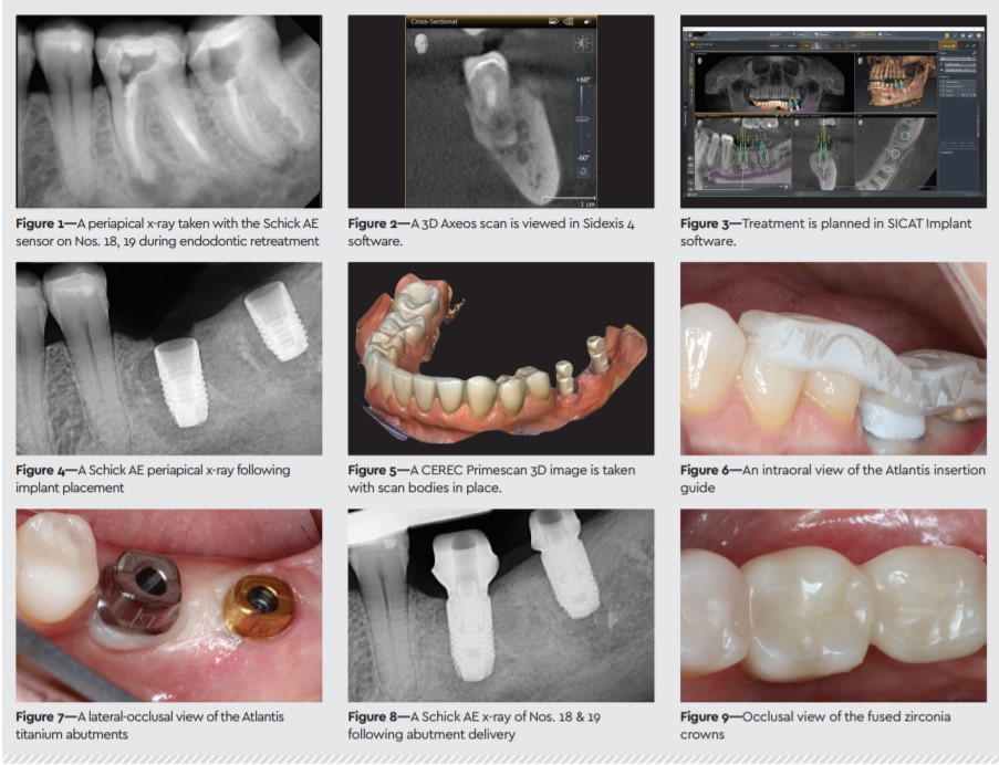
A 51-year-old female presented with continuous pain in the lower left jaw. She made me aware of past experiences including wisdom teeth removal complications where she needed extraoral incisions to drain an infection. Needless to say, this patient was very concerned upon learning that two of her endodontically treated teeth were abscessed and needed to be replaced. Complicating the situation was the extent of the infection, which spread to the inferior alveolar nerve canal apical to teeth Nos. 18 and 19 (Figure 1).
After the patient’s CBCT is acquired, I always invite the patient to view the 3D images with me. This allows the patient to understand the procedure better and take part in the planning process, while also facilitating informed consent. With the patient watching, I plan the preliminary implant position and explain how this is the safest and most predictable approach for implant placement. The patient is always impressed to see her teeth imported in an intraoral scan and superimposed with the 3D images. That’s a complete “wow” factor. In this case, the phobic patient understood that her assumed complicated surgery was very straightforward and she worried much less.
Treatment Workflow
After extraction, bone grafting, and subsequent healing, intraoral scans were taken of the upper and lower jaws using Dentsply Sirona’s CEREC Primescan, and the fi les were exported to SICAT Implant planning software. Here, the dentist can merge the 3D x-ray from Axeos with the 3D intraoral scans so accuracy and predictability can be guaranteed. After planning the implant positioning using Sidexis 4 and SICAT Implant 2.0 software (Figures 2–3), the dentist can design a surgical guide for printing in-house or by an outside provider, as was chosen in this case.
The guided surgery was accomplished without any complications (Figure 4). Two Astra Tech Implant System EV 4.8 implants were successfully delivered with transmucosal healing abutments. After an 8-week healing period, the successful osseointegration was checked with an Osstell IDx. Primescan was used to scan the implant position as well as scan bodies (Figure 5) so the Atlantis lab could fabricate custom abutments, which were shipped to our office. A core file of the Atlantis lab design allowed us to fabricate the crowns using our CEREC mill. The patient returned and, without the need for local anesthetic, the formerly anxious patient experienced a short and straightforward delivery appointment—and a beautiful outcome (Figures 7–9). This once-anxious patient even left us a wonderful online review. Meanwhile, the practice enjoyed a workflow that was predictable and, therefore, time- and money-saving.
AXEOS
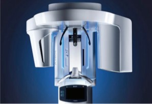
The Axeos 3D/2D imaging system uses intelligent low-dose exposure to capture high-quality images, while easy-to-use features, such as smart height adjustment and quick scan times, enhance patient comfort. Axeos is powered by Sidexis 4 imaging software and seamlessly integrates with over 250 practice management systems and multiple treatment planning software, such as SICAT Implant, SICAT Endo, SICAT Function, and SICAT Air. With the largest fi eld of view of any Dentsply Sirona 3D/2D imaging system, experience the difference Axeos can make in your practice.
Dr. Langenbach has provided his dental expertise to patients in both his native Germany and the United States. While serving as a dentist in the German Army, Dr. Langenbach met Dr. Lynne Thomas and they agreed that southern California was the place to be. Setting up their practice in Poway, CA, in 2003, Dr. Langenbach has consistently offered top-tier cosmetic, restorative, preventive dentistry, and sleep apnea treatments. Among his esteemed accreditations and affi liations, Dr. Langenbach is a Master of the Academy of General Dentistry and a Visiting Faculty for CDOCS. His main goal with any patient is to achieve beautiful, safe, and longstanding outcomes.