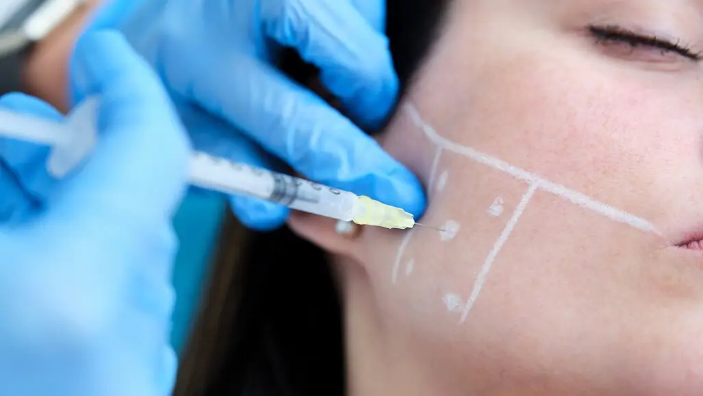
Hypertrophy of the masseter muscle is defined as the excessive growth of muscle mass in the transverse direction and is more common between the second and fourth decades of life. Even though its aetiology is multifactorial, bruxism is the main cause. There are many treatment alternatives, among which injection of botulinum toxin into the masseter muscle has been shown to be a safe and effective option.¹
Botulinum toxin is a powerful neurotoxin that, when injected into the muscle, generates interference with the neurotransmitter mechanism, inhibiting the release of acetylcholine at motor neuron axon terminals and thereby producing selective paralysis, decreased contraction and consequent muscular atrophy.² ³ It is this last effect that is exploited for addressing hypertrophy of the masseter, reducing its size and improving the patient’s aesthetics.
There is extensive literature on the parameters that must be followed for the injection of botulinum toxin to avoid risk areas and to reduce complications. In 2005, Kim et al. delineated a safe zone for the injection of botulinum toxin, demarcated vertically by the anterior and posterior boundaries of the masseter muscle and horizontally by a line drawn along the inferior border of the mandible to the mandibular angle and a line drawn from the base of the earlobe towards the corner of the mouth.⁴ In 2010, Kim et al. determined the subdivision of the masseter muscle into six zones, according to which injection into Zones I, II and III would be associated with adverse effects, such as injury to the parotid duct, and injection into Zone VI would not be effective for masseteric hypertrophy, establishing Zones IV and V as safe for botulinum toxin injection.⁵

Fig. 1a: Initial extra-oral photographs.

Fig. 1b: Initial extra-oral photographs.

Fig. 1c: Initial extra-oral photographs.
Case report
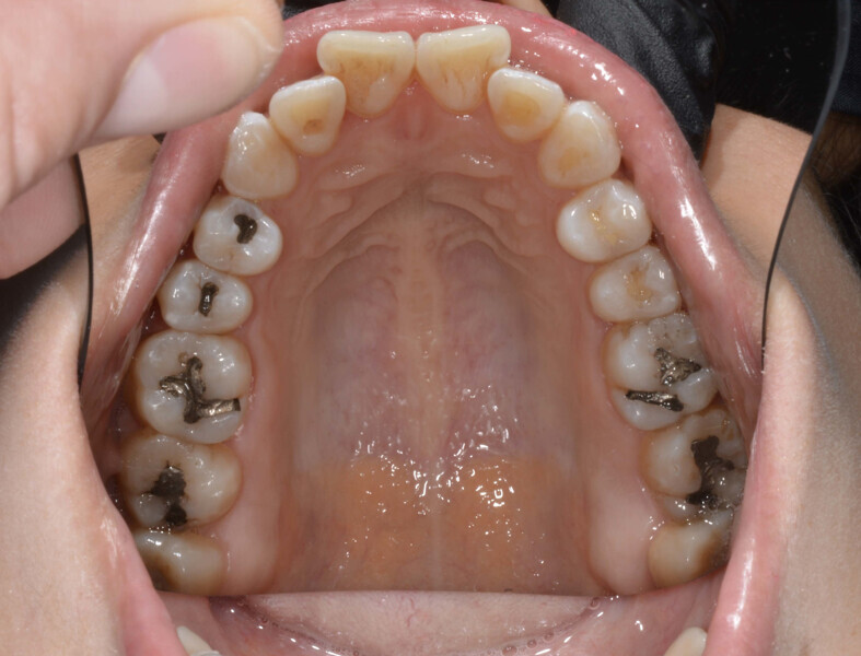
A 35-year-old female patient presented with crowding, muscle and joint pain, and headaches upon awakening. Examination found a skeletal Class I with crowding and bimaxillary protrusion. Facial examination showed a convex profile with protruded lips and a square face shape, intensified on smiling (Figs. 1–4). She reported bruxism, and bilateral masseteric hypertrophy was observed, but there was no alteration of the temporomandibular joints.