
Periapical x-rays are a foundational method in dental imaging. They are mainly designed to seize precise images of each the seen crown and the basis shape of a teeth, in addition to the encompassing bone. Many humans ask: Can a periapical x-ray sincerely display bone loss? Understanding the solution is crucial for everybody worried approximately their oral fitness or present process dental assessment.
What Is a Periapical X-Ray?
A periapical x-ray is a sort of dental radiograph that indicates the complete shape of a teeth, from the top (crown) all of the manner to the end of the basis. Importantly, it is usually the bone tissue that helps the teeth. Dentists use periapical x-rays due to the fact they offer clean pix of regions that can’t be visible all through a fashionable dental exam. These x-rays are regularly a part of a full-mouth series, which allows dentists examine basic oral fitness.
How Does Bone Loss Occur withinside the Jaw?
Bone loss withinside the jaw maximum generally effects from periodontal sickness, additionally referred to as gum sickness. The sickness starts offevolved as irritation of the gums, which, if untreated, can development deeper and begin to spoil the bone across the teeth. When bone is lost, the aid for the teeth weakens, that could in the end result in teeth mobility or maybe teeth loss. Detecting bone loss early is vital for powerful intervention and management.
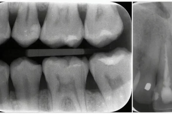
Can a Periapical X-Ray Show Bone Loss?
The easy solution is yes. Periapical x-rays are mainly used to come across bone loss round teeth. They offer pix displaying the bone degree adjoining to every teeth. This is crucial due to the fact bone loss underneath the gumline isn’t seen all through a everyday visible examination. The x-ray lets in the dentist to peer how plenty bone is assisting the teeth and to pick out early symptoms and symptoms of bone deterioration.
What Does Bone Loss Look Like on a Periapical X-Ray?
On a periapical radiograph, wholesome bone typically seems as a continuous, uniform shadow across the teeth root. When bone loss occurs, there are seen changes. The bone line appears much less even, seems decrease than regular, or can also additionally display angular defects. In mild or excessive cases, the bone assisting the basis can also additionally recede notably or seem much less dense.
Dentists are educated to identify those patterns. Experience and know-how make it feasible to differentiate regular anatomical versions from symptoms and symptoms of periodontal damage. However, sure anatomical capabilities and overlapping systems can every now and then make assessment challenging, mainly withinside the higher jaw wherein the sinuses or nasal cavities can also additionally difficult to understand key regions.
Diagnostic Accuracy and Limitations
Periapical x-rays are especially valued for his or her capacity to show hidden sickness. Studies have proven that each skilled dentists and synthetic intelligence fashions can come across bone loss with moderately excessive accuracy the use of periapical radiographs. In general, periapical x-rays supply diagnostic accuracy prices of over 80% for detecting periodontal bone loss.
However, no imaging technique is perfect. Some barriers exist. Image distortion can arise because of bad alignment of the x-ray or affected person movement. Overlapping anatomical structures, specifically withinside the higher jaw, may also difficult to understand element. In addition, early bone lack of minimum intensity or withinside the buccolingual direction (cheek-to-tongue side) might not be captured definitely on a fashionable 2D x-ray image. Severity of ailment and the precise web website online of bone destruction affect how a whole lot is seen.
Comparison with Other Imaging Techniques
While periapical x-rays provide top notch element for analyzing person enamel and their surrounding bone, different dental imaging modalities additionally exist. Bitewing x-rays are frequently used for detecting cavities and slight bone loss among returned enamel. Panoramic x-rays seize the complete jaw in a single image, however may also display overlapping tissues that difficult to understand element, specifically withinside the the front of the mouth. Cone beam computed tomography (CBCT) affords a three-D view and is now and again used while even extra element is needed, along with earlier than dental implant placement or for comparing complicated cases.
Despite those alternatives, periapical x-rays continue to be a cost-powerful and broadly on hand technique for the preliminary evaluation of bone fitness round enamel.
Technological Advances: Artificial Intelligence in Bone Loss Detection
Advanced studies has investigated the usage of synthetic intelligence, specifically convolutional neural networks (CNNs), to assist discover bone loss on periapical x-rays. While AI structures have proven promise, they acquire accuracy prices much like skilled dentists, frequently detecting bone loss with over 80% precision. Deep getting to know fashions advantage from massive, well-annotated information units, and the exceptional of those information units affects diagnostic performance.
AI can quick examine massive numbers of images, spotlight areas of concern, or even categorize the severity of bone loss. These technology assist standardize tests and can lessen human error. However, cautious affected person-precise assessment via way of means of a dental expert stays essential.
Clinical Relevance: Why Early Detection Matters
Bone loss is a silent procedure in its early stages. Patients frequently enjoy no ache or seen signs till widespread harm has occurred. By the time a teeth will become free or a pocket paperwork withinside the gum, great bone loss may also have already taken place. Early detection the usage of periapical x-rays permits dentists to interfere quick.
Timely remedy may also encompass stepped forward oral hygiene, expert cleaning, or extra superior periodontal therapies. By figuring out bone loss early, dentists can assist save you the want for teeth extraction and keep long-time period oral fitness and function.
The Periapical X-Ray Experience: What Patients Should Expect
Having a periapical x-ray taken is a brief and normally snug procedure. Patients are seated in a dental chair and guarded with a lead apron. The x-ray movie or virtual sensor is located withinside the mouth the use of a holder. The dentist or hygienist aligns the x-ray machine, and the publicity lasts most effective a short moment. Modern dental x-rays use very low doses of radiation, making them secure for recurring use while justified via way of means of scientific need.
After the picture is taken, it’s far reviewed for first-rate to make sure all regions of hobby are seen and actually represented. If something is doubtful or if anatomical versions difficult to understand the view, a repeat x-ray can be had to enhance diagnostic accuracy.
Conclusion
Periapical x-rays are a crucial diagnostic device for detecting bone loss in dentistry. They offer clean pics of the teeth shape and the helping bone, permitting dentists to discover issues that could in any other case continue to be hidden. Bone loss may be actually visualized on those radiographs, specifically while it has reached a positive diploma of severity. While a few technical limits exist, periapical x-rays are fantastically powerful and secure for recurring use. Dentists rely upon them to manual remedy making plans and interventions for preserving lifelong oral health.
FAQs
Can a periapical x-ray come across early bone loss?
Yes. Periapical x-rays can display early symptoms and symptoms of bone loss, despite the fact that very minor modifications can be greater tough to come across. Their sensitivity improves as the quantity of bone loss increases.
Are periapical x-rays secure?
Yes. Modern dental x-rays are very secure. They use minimum radiation and are most effective endorsed while clinically necessary.
How frequently ought to I even have a periapical x-ray?
Frequency relies upon for your dental health, chance factors, and your dentist’s recommendations. They are frequently carried out as a part of a diagnostic technique while gum disorder or different issues are suspected.
What ought to I do if bone loss is determined on my x-ray?
Follow your dentist’s advice, which might also additionally consist of expert cleaning, advanced oral hygiene, or referral to a periodontal specialist.
Can kids have bone loss detected with periapical x-rays?
Yes. While bone loss is much less not unusualplace in kids, periapical x-rays can display gum disorder or different bone modifications if present.