


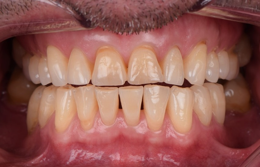
What Is a Wedge-Shaped Defect?
A wedge-shaped defect is a loss of hard tooth tissue near the neck of a tooth.
The neck is the area close to the gumline. Dentists call this region the cervical area of the tooth.
The defect often appears as a V-shaped notch. Its shape may resemble a carpenter’s wedge. Therefore, it is commonly called a wedge-shaped defect in patient education.
However, dentists often use a broader clinical term:
Non-carious cervical lesion, or NCCL.
“Non-carious” means the tissue loss does not start from bacterial tooth decay. “Cervical” refers to the neck of the tooth. “Lesion” describes the damaged area.
Not every cervical lesion has a sharp V shape.
Some defects appear:
- Narrow and sharply angled
- Broad and shallow
- Smooth and saucer-shaped
- Irregular or asymmetrical
- Polished and glossy
- Rough near the deepest area
Therefore, the visible shape alone does not always reveal the exact cause.
In addition, more than one type of wear may occur at the same time.
For example, acid may soften the tooth surface. Then, forceful brushing may remove the softened tissue more easily.
Modern research generally treats these lesions as multifactorial. Excessive brushing and acidic exposure are common contributors. Meanwhile, the role of bite-related tooth flexure remains debated.
Brushing Is Simple, but Brushing Incorrectly Can Damage Teeth
Many people see brushing as a simple daily task.
However, brushing technique can greatly affect oral health. Correct brushing removes plaque and food debris from the teeth and gumline. Therefore, it helps reduce the risk of tooth decay and gum disease.
In contrast, excessive or incorrect brushing may damage the teeth and gums.
Some people often experience tooth sensitivity or a short, sharp pain. Cold water may trigger it. Cold air, sweet foods, or acidic drinks may also cause discomfort.
As a result, many patients assume that they have tooth decay.
However, decay is not the only possible cause.
A wedge-shaped defect near the gumline can also expose dentin. Once the dentin becomes exposed, cold, heat, touch, and acidic foods may trigger sensitivity.
This type of damage usually develops slowly. Therefore, many people do not notice it during the early stage.
They may only seek dental care when the defect becomes deeper or starts to hurt.
Although aggressive horizontal brushing can contribute to the problem, it is rarely the only factor. Instead, several chemical and mechanical forces often act together.
For this reason, treatment should not focus only on changing the brushing direction. The dentist must also identify other possible causes.
Where Do These Defects Usually Appear?
Wedge-shaped cervical defects usually form near the gumline.
They often affect the outer surfaces of the teeth. These are the surfaces next to the cheeks or lips.
Premolars and canines commonly show visible cervical wear. These teeth sit near the curved areas of the dental arch.
As a result, they may receive more brushing pressure. They may also become more exposed after gum recession.
However, any tooth can develop a cervical lesion.
The location depends on several factors:
- The person’s brushing habits
- Toothbrush stiffness
- Hand dominance
- Gum position
- Tooth alignment
- Acid exposure
- Tooth structure
- Bite forces
- Existing dental restorations
Some patients show similar defects on both sides of the mouth.
Others have more damage on one side. For example, a right-handed person may brush the left side more aggressively.
Still, this pattern is not reliable enough to identify the cause by itself.
A dentist must examine the entire mouth. The dentist may also ask about diet, brushing, acid reflux, tooth grinding, and previous dental treatment.
The location offers useful clues. However, it rarely gives the complete answer.
What Does It Feel Like?
A mild defect may cause no symptoms.
Therefore, a person may not know that any tooth tissue has been lost.
As the lesion becomes deeper, it may expose dentin. Dentin lies beneath the harder enamel layer.
Dentin contains tiny channels that connect toward the inner pulp. When cold, air, pressure, or acidic substances affect these channels, the tooth may feel sensitive.
Common symptoms include:
- A short, sharp pain from cold water
- Sensitivity when breathing cold air
- Discomfort when brushing the gumline
- Pain from sweet or sour foods
- Sensitivity when touching the defect
- An uneven or indented area near the gum
- Food or plaque collecting around the lesion
The pain often disappears soon after the trigger stops.
However, deeper damage may cause stronger or longer discomfort.
Persistent pain may suggest another problem. For example, the tooth could have decay, a crack, pulp inflammation, or gum disease.
Therefore, patients should not diagnose the condition only from the symptoms.
A dental examination helps separate a cervical defect from decay and other dental conditions.
Cause One: Incorrect Brushing Technique
Incorrect brushing is an important risk factor.
Long-term horizontal scrubbing may repeatedly rub the cervical area. Excessive force can make this effect stronger.
A hard toothbrush may also increase mechanical wear.
However, the direction of brushing is not the only concern.
A person may use a vertical or circular method but still press too hard. Likewise, a soft brush can cause wear when used aggressively for a long time.
Several brushing habits may increase the risk:
- Using strong hand pressure
- Scrubbing with long horizontal strokes
- Using a medium or hard toothbrush
- Brushing one area for too long
- Replacing gentle movement with rapid sawing
- Using a highly abrasive toothpaste
- Brushing immediately after acidic food or vomiting
- Using an old brush with damaged bristles
Therefore, prevention requires more than changing from horizontal to vertical brushing.
The person should reduce pressure and use controlled movements.
An electric toothbrush with a pressure sensor may help some patients. However, it can still cause irritation if used incorrectly.
The main goal is to remove plaque without repeatedly grinding the tooth surface.
Why Brushing Alone Does Not Explain Every Case
For many years, people linked cervical defects mainly to aggressive toothbrushing.
However, brushing alone cannot explain every clinical pattern.
Some patients develop lesions even though they use a soft toothbrush. Others brush forcefully but show little cervical damage.
In addition, the lesions may occur on teeth that receive limited direct brushing.
This suggests that several factors can work together.
For example:
- Acid softens the tooth surface.
- Gum recession exposes less protected root tissue.
- Brushing removes part of the softened surface.
- Repeated bite forces may concentrate stress near the neck.
- The lesion slowly becomes deeper.
The exact combination differs between patients.
Therefore, dentists should avoid blaming the patient’s brushing without further assessment.
Instead, the dentist should ask:
- How often does the patient consume acidic drinks?
- Does the patient have heartburn or reflux?
- Is gum recession present?
- Does the patient grind or clench?
- Which toothpaste does the patient use?
- How soon after meals does the patient brush?
- Is the lesion changing over time?
This broader evaluation leads to more effective prevention and treatment.
Cause Two: The Cervical Area Has Less Protection
The neck of a tooth has a special structure.
The crown of the tooth has a thick enamel covering. Enamel is the hardest tissue in the human body.
However, the enamel becomes thinner near the gumline.
Below this area, the root surface has a different covering called cementum. Cementum is much softer than enamel.
When the gums stay healthy and cover the root, this softer tissue remains protected.
However, gum recession may expose it.
Once exposed, the root surface can wear more quickly. It can also become sensitive.
Several conditions may contribute to gum recession:
- Aggressive brushing
- Gum inflammation
- Periodontal disease
- Naturally thin gum tissue
- Tooth position outside the dental arch
- Orthodontic movement
- Aging
- Smoking
- Oral piercings
- Previous gum surgery
Therefore, cervical damage may involve both the tooth and the surrounding gums.
Simply filling the notch may not solve the full problem.
The dentist must also check whether the gums continue to recede.
In some cases, periodontal treatment or gum grafting may become part of the long-term plan.
Cause Three: Toothbrush Pressure from the Cheeks and Lips
During brushing, the cheeks and lips may press against the toothbrush.
This contact can increase pressure on the outer tooth surfaces.
However, cheek pressure is not usually considered an independent primary cause of a cervical lesion.
Instead, it may influence the way a person holds and moves the brush.
For example, the curved part of the dental arch may receive greater contact. The toothbrush may also stay at an incorrect angle in that area.
As a result, certain teeth may receive repeated friction.
Canines and premolars often sit near this curve. Therefore, they commonly show cervical wear.
Still, the exact tooth pattern differs from person to person.
The important point is not the cheek muscle alone. The main issue is repeated mechanical force concentrated on a small cervical area.
A dentist or hygienist can demonstrate a safer brushing angle.
The bristles should reach the gumline gently. However, they should not bend strongly against the teeth.
Small, controlled movements usually work better than forceful scrubbing.
Cause Four: Acid Exposure
Acid plays an important role in many non-carious cervical lesions.
Acid can soften enamel and dentin. Once the surface becomes softer, brushing and chewing may remove it more easily.
Acid may come from outside or inside the body.
Common external sources include:
- Soft drinks
- Sports drinks
- Energy drinks
- Citrus fruit
- Fruit juice
- Vinegar
- Sour candy
- Carbonated water with added acid
- Frequent wine consumption
- Acidic vitamin drinks
Internal acid usually comes from the stomach.
Possible causes include:
- Gastroesophageal reflux
- Frequent heartburn
- Repeated vomiting
- Eating disorders
- Pregnancy-related vomiting
- Certain digestive conditions
- Heavy alcohol use
A person with reflux may not always feel obvious heartburn. Therefore, dental wear can sometimes provide an early clue.
However, not every cervical lesion comes from acid.
The dentist looks at the location and appearance of wear. Acid erosion often affects several surfaces and may create smooth, rounded areas.
When acid and brushing occur together, the pattern may become more localized.
Should You Brush Immediately After Acid Exposure?
Brushing immediately after an acidic drink or vomiting may increase surface wear.
The acid temporarily softens the outer tooth tissue.
Therefore, forceful brushing during this period may remove more tissue.
After acidic food or drink, a person can:
- Rinse the mouth with plain water
- Drink water
- Avoid holding the drink in the mouth
- Use a straw when appropriate
- Wait before brushing
- Chew sugar-free gum if suitable
- Reduce the frequency of acidic intake
After vomiting or reflux, the person should rinse gently.
They should not immediately scrub the teeth.
However, delaying brushing does not mean leaving plaque on the teeth for many hours.
The main focus should be reducing frequent acid exposure and avoiding strong brushing on softened surfaces.
Patients with repeated reflux should seek medical advice.
Dental treatment alone cannot control stomach acid.
Cause Five: Bite Stress and Tooth Flexure
Some dental theories suggest that heavy bite forces can contribute to cervical defects.
During clenching or grinding, a tooth may flex very slightly.
Supporters of this theory suggest that stress becomes concentrated near the tooth neck. Over time, tiny changes in the hard tissue may occur.
This proposed process is often called abfraction.
However, the role of abfraction remains controversial.
Research has not proven that bite stress alone creates most wedge-shaped lesions. Some reviews describe it as a possible contributing factor rather than a confirmed single cause.
Therefore, dentists should not automatically adjust the bite or prescribe a night guard only because a cervical notch exists.
They should first look for other signs, such as:
- Tooth grinding
- Flattened biting surfaces
- Cracks
- Jaw muscle pain
- Broken restorations
- Tooth mobility
- Heavy bite contacts
- Morning jaw fatigue
When these signs exist, managing grinding may help protect the teeth.
However, bite treatment should follow a complete diagnosis.
Unnecessary removal of tooth structure through bite adjustment may create new problems.
Are Bacteria the Main Cause?
Bacteria cause tooth decay by producing acids from sugars.
However, a wedge-shaped non-carious cervical defect is not primarily a bacterial disease.
This distinction is important.
Dental plaque may collect inside an existing defect. The rough or indented area can be harder to clean.
As a result, decay may later develop on top of a non-carious lesion.
Still, bacteria did not necessarily create the original notch.
Therefore, dentists must distinguish between:
- A non-carious cervical lesion
- Root surface decay
- A combination of cervical wear and decay
The color and texture may offer clues.
A non-carious lesion often feels hard and smooth. Active decay may feel softer or rougher.
However, visual inspection alone is not always enough.
The dentist may dry the area, examine it carefully, check sensitivity, and use X-rays when necessary.
Calling every cervical defect “decay” can lead to the wrong treatment.
Likewise, assuming every notch is harmless wear may delay necessary caries treatment.
How Dentists Diagnose a Cervical Defect
A dentist begins with a clinical examination.
The defect’s position, shape, depth, color, and surface texture provide useful information.
The dentist may also ask when the sensitivity started and what triggers it.
Important questions may include:
- Which toothbrush do you use?
- How much pressure do you apply?
- Which toothpaste do you use?
- How often do you consume acidic foods?
- Do you drink soft drinks slowly throughout the day?
- Do you experience reflux?
- Do you grind your teeth?
- Has the gumline changed?
- Is the defect becoming larger?
- Does the pain continue after the trigger stops?
The dentist may take photographs or digital scans.
These records help monitor the lesion.
In addition, the dentist may measure the depth or mark the defect on a dental chart.
X-rays may help rule out decay or pulp problems. However, a shallow cervical lesion may not appear clearly on an X-ray.
The diagnosis often depends on combining several findings.
Treatment for a Mild Defect
A small lesion without sensitivity may not need immediate filling.
Instead, the dentist may monitor it.
This approach works best when:
- The defect is shallow
- The surface remains hard
- The lesion is not progressing
- The tooth has no sensitivity
- Food does not collect in it
- The tooth remains strong
- The appearance does not concern the patient
However, monitoring should remain active.
The dentist may take photographs or measurements during regular visits.
At the same time, the patient should reduce the risk factors.
Recommended changes may include:
- Using a soft toothbrush
- Reducing brushing pressure
- Choosing a less abrasive toothpaste
- Avoiding repeated horizontal scrubbing
- Reducing acidic drinks
- Managing reflux
- Using fluoride products
- Treating dry mouth
- Avoiding brushing immediately after acid exposure
Changing the causes may slow or stop progression.
In contrast, filling a lesion without changing the causes may lead to continued wear around the restoration.
How to Manage Tooth Sensitivity
Sensitivity does not always mean that the tooth needs a filling.
For a mild lesion, the dentist may first recommend non-invasive care.
Options may include:
- Desensitizing toothpaste
- Fluoride varnish
- High-fluoride products when appropriate
- Dentin-sealing agents
- Resin-based desensitizers
- Improved brushing technique
- Reduced acid exposure
Desensitizing toothpaste usually needs regular use.
The patient should not expect one application to solve the problem immediately.
Some products work by blocking the tiny channels in dentin. Others reduce nerve response.
The patient can place a small amount of toothpaste on the sensitive area after brushing. However, they should follow the product instructions or the dentist’s advice.
If sensitivity continues, the dentist should reassess the tooth.
Long-lasting pain may suggest a crack, decay, pulp inflammation, or another condition.
Therefore, repeated self-treatment without examination may hide a more serious problem.
Treatment for a Moderate Defect
A moderate lesion may need restoration.
The dentist may recommend filling when:
- The defect continues to deepen
- Sensitivity affects daily life
- The shape traps plaque or food
- The tooth looks weak
- The patient dislikes the appearance
- The lesion lies near a denture or appliance
- The remaining tooth tissue may fracture
- The defect makes brushing difficult
Common restorative materials include composite resin and glass ionomer materials.
Composite resin can closely match the tooth color. It also allows the dentist to shape and polish the area.
Glass ionomer can release fluoride and may bond well in selected cervical areas.
Some dentists use resin-modified glass ionomer. Others combine materials depending on the lesion and moisture control.
No single material suits every case.
The dentist considers gum position, isolation, bite stress, sensitivity, appearance, and lesion depth.
What Happens During the Filling Procedure?
The dentist first cleans the lesion.
Local anesthesia may not always be necessary. However, it may help when the tooth is sensitive or the defect is deep.
The dentist may lightly prepare the surface. In many cases, extensive drilling is unnecessary.
Next, the area must remain as clean and dry as possible.
The dentist applies the selected bonding system and restorative material.
Then, the material is shaped to follow the natural contour of the tooth.
The dentist also checks the bite.
A cervical filling should not create a bulky edge near the gumline. Otherwise, it may collect plaque or irritate the gums.
Finally, the restoration is polished.
The patient should continue using gentle brushing after treatment.
A filling repairs the lost shape. However, it does not remove the cause.
Therefore, strong brushing, acid exposure, or other risk factors can still damage the surrounding tooth or restoration.
Treatment for a Severe Defect
A deep wedge-shaped defect can weaken the tooth.
In severe cases, the lesion may approach the dental pulp.
The patient may experience strong sensitivity or pain.
If the pulp remains healthy, the dentist may protect it and restore the lost structure.
However, pulp inflammation may develop when the defect becomes very deep or when cracks and bacteria affect the inner tissue.
Possible warning signs include:
- Spontaneous toothache
- Pain that wakes the patient at night
- Lingering pain after cold or heat
- Pain when biting
- Swelling
- Gum tenderness
- Tooth discoloration
- A draining gum abscess
These symptoms require prompt dental care.
If irreversible pulp inflammation occurs, root canal treatment may be necessary.
If infection spreads beyond the root, the dentist must also treat the root-end tissues.
After root canal treatment, the tooth may need a strong final restoration.
In extreme cases, a severely weakened tooth may fracture and become difficult to save.
Therefore, early assessment can prevent a manageable lesion from becoming a complex dental problem.
Can Gum Treatment Be Necessary?
Some cervical defects occur together with gum recession.
In these cases, the dentist may consider both restorative and periodontal treatment.
A filling can cover exposed dentin. However, it cannot always restore the lost gumline.
For selected patients, gum grafting may help cover part of the exposed root.
The treatment choice depends on:
- The amount of gum recession
- Gum thickness
- Root surface condition
- Lesion depth
- Tooth position
- Smile appearance
- Plaque control
- Patient preference
Sometimes the dentist restores part of the defect and a periodontist treats the gum recession.
In other cases, the gum procedure comes first.
However, not every recessed area requires surgery.
The dentist should explain the expected result and limitations.
The goal is to improve comfort, protect the tooth, support gum health, and create a cleanable surface.
Can a Wedge-Shaped Defect Be Prevented?
Many cervical lesions can be reduced or slowed.
However, prevention requires control of several factors.
First, choose a soft-bristled toothbrush.
Second, use gentle pressure.
The bristles should touch the teeth and gumline. However, they should not flatten strongly.
Third, avoid forceful back-and-forth scrubbing.
Small circular or short controlled strokes usually provide better control.
A modified Bass technique may also help. In this method, the bristles sit gently near the gumline and move with small vibrations.
However, no single technique suits every patient.
People with braces, gum disease, limited hand movement, or dental restorations may need personalized guidance.
Therefore, ask a dentist or hygienist to demonstrate the method.
Technique matters more than simply calling the movement “vertical” or “horizontal.”
Choose Toothpaste Carefully
Toothpaste helps remove plaque and deliver fluoride.
However, toothpaste also contains abrasive particles.
These particles help remove stains and debris. Yet very abrasive products may increase wear when combined with heavy brushing.
Whitening toothpastes can vary widely in abrasiveness.
Charcoal products may also have uncertain or inconsistent abrasive effects.
Therefore, patients with cervical wear should avoid choosing toothpaste only by its whitening claim.
A fluoride toothpaste with a mild formula may suit sensitive teeth better.
Patients can look for products designed for:
- Sensitive teeth
- Enamel protection
- Gentle cleaning
- Low-abrasion care
However, labels do not always provide the full abrasion level.
A dentist can recommend a suitable product.
In addition, using more toothpaste does not improve cleaning.
A pea-sized amount is enough for most adults.
The brushing method and duration matter more than a large amount of paste.
Control Acidic Foods and Drinks
Patients do not need to avoid every acidic food.
Instead, they should reduce frequent and prolonged exposure.
Frequency often matters more than the total amount.
For example, slowly sipping a soft drink for several hours exposes the teeth repeatedly.
Drinking it with a meal creates a shorter exposure.
Helpful habits include:
- Drink acidic beverages less often
- Avoid holding drinks in the mouth
- Do not swish soft drinks around the teeth
- Use water as the main daily drink
- Rinse with water after acidic intake
- Avoid frequent sour candy
- Do not sip lemon water all day
- Discuss ongoing reflux with a doctor
- Avoid brushing immediately after vomiting
Patients should also remember that “sugar-free” does not always mean “acid-free.”
A sugar-free carbonated drink may still contribute to erosive wear.
Therefore, checking acidity matters as well as checking sugar.
Regular Self-Checks Can Help
A person can check the visible tooth surfaces in a mirror.
Good lighting can make the gumline easier to see.
Possible warning signs include:
- A small notch near the gum
- A yellow or darker area at the tooth neck
- A shiny depression
- A rough edge
- Gum recession
- Sensitivity during brushing
- A defect that appears larger over time
However, home inspection has limits.
Early lesions can be difficult to see. They may also hide behind the lips or between teeth.
In addition, a dark area may represent a stain, exposed dentin, decay, or an old restoration.
Therefore, self-checking cannot replace dental examinations.
It simply helps patients notice changes earlier.
Photographs can also help. A patient may take a clear image under similar lighting every few months.
Still, a dentist should assess any suspected defect.
When Should You See a Dentist?
Do not wait until the tooth becomes severely painful.
Arrange a dental examination when:
- Cold sensitivity happens repeatedly
- The gumline looks indented
- A notch becomes larger
- The tooth hurts during brushing
- Food collects in the defect
- The gum starts to recede
- The tooth looks cracked
- Pain continues after the trigger stops
- The tooth hurts without a clear trigger
- Swelling develops
Early care often requires fewer procedures.
For example, a shallow lesion may only need risk-factor control and desensitizing care.
A moderate lesion may need a small restoration.
In contrast, a deep lesion may require root canal treatment or more extensive reconstruction.
Therefore, early detection can protect more natural tooth tissue.
Common Misunderstandings
One misunderstanding is that horizontal brushing causes every cervical notch.
In fact, aggressive brushing may contribute, but acid exposure, gum recession, tissue structure, and other forces may also matter.
Another misunderstanding is that every defect must be filled immediately.
A shallow and stable lesion may only need monitoring.
Some people also believe that sensitivity always means tooth decay.
However, exposed dentin can also cause sharp pain.
Another belief is that bacteria directly create all wedge-shaped defects.
Non-carious cervical lesions do not begin in the same way as cavities. Still, decay may later develop inside them.
Finally, some patients believe that a filling permanently solves the problem.
A restoration replaces lost tissue. However, the lesion may continue around it if brushing pressure or acid exposure remains unchanged.
Therefore, successful care combines treatment with prevention.
Frequently Asked Questions
Is a Wedge-Shaped Defect the Same as a Cavity?
No. A wedge-shaped cervical defect usually involves non-carious tooth wear. However, decay can sometimes develop in the same area.
Can Brushing Too Hard Cause It?
Yes, excessive brushing may contribute. However, it often works together with acid exposure, gum recession, or other factors.
Does Every Cervical Defect Need a Filling?
No. A small, stable lesion without symptoms may only need monitoring and preventive care.
Can the Lost Tooth Structure Grow Back?
No. Lost enamel and dentin do not grow back naturally. However, dentists can control sensitivity and restore deeper defects.
Which Toothbrush Is Best?
A soft-bristled toothbrush usually works well. More importantly, use gentle pressure and avoid forceful scrubbing.
Summary
A wedge-shaped defect is a form of tooth tissue loss near the gumline.
It may appear as a sharp V-shaped notch. However, some lesions look broad, smooth, or shallow.
Aggressive horizontal brushing can contribute to the problem. Still, it is not the only possible cause.
Acidic drinks, gastric reflux, exposed root surfaces, gum recession, abrasive toothpaste, and repeated mechanical forces may also play a role.
Meanwhile, the effect of bite-related tooth flexure remains controversial. Bacteria are not considered the main direct cause of a non-carious lesion.
Treatment depends on the depth, symptoms, progression, appearance, and strength of the tooth.
A mild lesion may need monitoring and preventive care. A sensitive lesion may respond to fluoride or desensitizing treatment. A deeper defect may need composite resin or glass ionomer restoration.
Severe damage may affect the dental pulp and require root canal treatment.
Therefore, do not wait for strong pain.
Check the gumline regularly and attend dental examinations. In addition, use a soft toothbrush, gentle pressure, a suitable toothpaste, and a controlled brushing method.
Most importantly, remember that brushing harder does not mean brushing better.
Good prevention comes from gentle cleaning, lower acid exposure, early detection, and treatment based on the true cause.