
Dental X-rays play a critical function in contemporary-day oral fitness care. Among the distinctive varieties of photographs utilized by dentists, bitewing and periapical X-rays are of the maximum important. Although they may appear just like patients, those X-ray strategies serve distinctive functions and offer particular insights into the fitness of tooth and surrounding systems. Understanding the distinction among a bitewing photograph and a periapical photograph let you recognize their precise price for diagnosing dental troubles and making plans powerful treatment.
What Is a Dental X-ray?
A dental X-ray is an imaging method that makes use of small quantities of radiation to create photos of the tooth, jaw, and surrounding tissues. These photographs can monitor troubles that aren’t seen at some stage in a general dental examination. X-rays are secure while executed efficiently and provide vital statistics for preventing, diagnosing, and coping with oral conditions. Today, virtual X-rays are common, imparting excessive high-satisfactory photographs with minimum radiation exposure.
What Is a Bitewing Image?
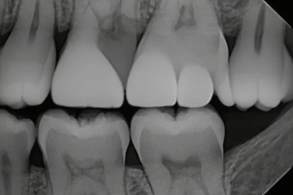
A bitewing photograph is a unique form of dental X-ray designed to expose info of the top and decrease returned tooth in a single area—typically the premolars and molars. The name “bitewing” comes from the wing-fashioned tool the affected person bites directly to keep the X-ray movie or sensor in place. The movie is placed in order that the X-ray will seize each the crowns of the top and decrease tooth and the bone among them.
Bitewing X-rays are especially used to:
- Detect cavities (dental caries) among tooth, which might be frequently hidden from view
- Monitor bone levels, specially for symptoms and symptoms of gum sickness (periodontitis)
- Check the suit and integrity of dental restorations like fillings or crowns
- Track any modifications to tooth and bone over time
Dentists normally use bitewing photographs at some stage in habitual dental checkups. These photographs are especially powerful in figuring out early teeth decay or modifications in bone density, which might be key signs of oral fitness troubles.
What Is a Periapical Image?
A periapical photograph is every other form of dental X-ray, however it specializes in a unmarried teeth (or some tooth) from the crown to the cease of the foundation and consists of the encircling bone. To create a periapical photograph, a sensor or movie is located at the back of the teeth and an X-ray beam is directed to seize all vital systems, which include the tip (apex) of the foundation.
Periapical X-rays are used to:
- Diagnose infections, abscesses, or different troubles across the root and surrounding bone
- Identify bone loss because of sickness or trauma
- Evaluate the quantity of harm after an injury
- Monitor the development of remedies consisting of root canals
- Detect peculiar growths or cysts close to the foundation
Dentists typically order periapical photographs while a affected person complains of ache or swelling in a selected area, or each time an in depth have a take a observe a teeth and its underlying systems is necessary.
The Key Differences: Bitewing vs Periapical Images
Anatomy Captured
Bitewing pictures display the crowns of each top and decrease enamel in a specific section, plus the peak of the bone among those enamel. The root suggestions and surrounding jawbone aren’t completely seen in bitewing pictures.
Periapical pictures display the whole tooth—from the crown right all the way down to the end of the foundation—and the bone shape across the root. These pictures aren’t as correct at shooting the touch region among adjoining crowns as bitewings are.
Diagnostic Focus
Bitewing pictures are exceptional for detecting cavities among enamel and tracking bone stages that is probably stricken by gum disease. Periapical pictures excel at highlighting issues that arise at the foundation or beneath the gum line, including abscesses, cysts, or intense bone loss.
Image Technique and Angulation
To take a bitewing photograph, the X-ray beam is placed at a particularly low vertical angulation (normally around +five stages). This angulation lets in for a clean view of the crown margins and call factors among adjoining enamel. Bitewings hold those edges separate, supporting dentists test for cavities and compare the match of fillings or crowns.
In contrast, periapical pictures normally require a extra vertical angulation (from +10 to +30 stages for top enamel) to seize the foundation suggestions. However, this better attitude can purpose the seen a part of the crown to overlap with the foundation, from time to time making it tough to peer the crown margin clearly. That method periapical X-rays aren’t the exceptional desire for locating cavities on the crown facet or for checking the bone stage simply beneath the crown.
Usefulness in Clinical Situations
- For checking for cavities among enamel and assessing the match of a brand new crown, a bitewing photograph is normally superior. The approach continues the crown margins and helping bone seen, warding off any overlaps that could conceal a problem.
- For diagnosing infections, tracking root canal treatments, or investigating ache localized in a single tooth, a periapical photograph is necessary, because it offers a whole view of the tooth’s roots and the bone wherein they’re anchored.
- In sufferers with sure anatomical features (including a shallow palate), periapical pictures can be tough to seize correctly. Sometimes the sensor can’t be parallel to the tooth, and the ensuing photograph can be foreshortened. In those cases, vertical bitewing pictures may be used as an opportunity to visualise the region properly.
Limitations and Best Practices
Bitewing pictures are restricted of their scope, as they do now no longer seize the foundation suggestions. They can omit infections or different root-associated issues. Periapical pictures, despite the fact that top notch for root detail, could make it tough to decide the touch factors or margins at the crown because of their angulation. For the exceptional dental care, dentists frequently use each kinds together, choosing the proper photograph primarily based totally at the medical question.
Which X-ray Does Your Dentist Choose?
Your dentist will determine among a bitewing or periapical photo relying for your signs and symptoms and the precise a part of your mouth that calls for assessment. Routine assessments typically depend upon bitewings to discover early cavities and display bone fitness. If you’ve got got unexplained pain, swelling, or a dental contamination is suspected, a periapical X-ray will probably offer the solutions your dentist needs. Sometimes, each pics are taken on the identical go to for an entire expertise of your dental fitness.
Conclusion
Bitewing and periapical X-ray pics are each critical equipment in dental diagnosis, however they’re designed to reply exceptional questions. Bitewing pics excel at locating cavities among enamel and tracking gum and bone fitness close to the crown. Periapical pics are higher for investigating deeper troubles associated with the roots or bone across the tooth. By expertise those differences, sufferers can higher recognize their dentist’s selections and sense assured of their oral care plan.
Frequently Asked Questions (FAQs)
1. Are Bitewing or Periapical X-rays More Painful?
Both forms of X-rays are typically brief and painless. There can be moderate soreness from protecting the sensor, however it lasts most effective a moment.
2. Is Radiation Exposure a Concern with These Dental X-rays?
Dental X-rays use very low doses of radiation. Modern structures use virtual sensors, which drastically lessen publicity in comparison to the past.
3. How Often Do I Need Each Type of X-ray?
The frequency relies upon for your oral fitness status. Bitewing pics are ordinary at normal checkups (yearly or biannually). Periapical X-rays are taken as wished if precise problems arise.
4. Can These X-ray Images Detect All Types of Dental Problems?
No unmarried kind can stumble on each problem. Bitewing pics are pleasant for cavities and bone levels. Periapical pics excel at root and bone problems. Dentists pick out the sort primarily based totally on what they want to diagnose.
5. Should I Be Worried About the Procedure?
There is not anything to fear about. Dental X-rays are ordinary and safe. If you’ve got got concerns, your dental crew will give an explanation for the system and cope with any questions you’ve got got.