
Alveolar bone loss is a essential component in dental and periodontal health, affecting each the stableness of enamel and the general circumstance of the mouth. Early and correct size of alveolar bone loss is important in diagnosing periodontitis, making plans dental remedies, and tracking the fulfillment of interventions. Understanding the cutting-edge techniques to quantify this bone loss can manual sufferers and clinicians in making knowledgeable choices for oral health.
What Is Alveolar Bone Loss?
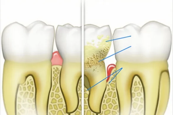
Alveolar bone is the bone that surrounds and helps your enamel. When this bone is lost, regularly because of periodontal (gum) disorder, enamel can grow to be unfastened and might ultimately fall out. Alveolar bone loss refers back to the discount in top or extent of this bone housing, commonly because of continual irritation. It is a number one marker for the severity of periodontitis and may sign the distinction among reversible and irreversible gum disorder.
Why Measure Alveolar Bone Loss?
Accurate size of alveolar bone loss is important for numerous reasons. Firstly, it allows in early prognosis of periodontal diseases. Secondly, it permits the dentist to evaluate the development of disorder and the effectiveness of treatment. Thirdly, it’s far an crucial step in making plans interventions like dental implants or crown lengthening. Finally, tracking bone loss can function a preventive degree, probably heading off enamel loss and extra invasive remedies down the line.
Traditional Methods for Measuring Alveolar Bone Loss
Visual Evaluation and Probing
Visual exam and probing are some of the oldest strategies utilized by clinicians. Dentists use a periodontal probe to degree the intensity of wallet across the enamel and to estimate wherein the bone crest lies underneath the gums. While this approach is simple, it’s far subjective and may be stricken by elements like irritation or probe angulation.
Use of Radiographs
Conventional dental X-rays (radiographs), which includes bitewing, periapical, and panoramic pix, had been the staple for assessing alveolar bone levels. Dentists visually look into those pix to estimate the gap from the cemento-tooth junction (CEJ) to the alveolar bone crest (ABC).
- Bitewing Radiographs: Best for viewing the top and decrease returned enamel and the bone round them. Frequently used to screen bone adjustments over time.
- Periapical Radiographs: Capture the total enamel duration and surrounding bone, top for assessing localized bone loss.
- Panoramic Radiographs: Provide a top level view of the enamel and jaws, beneficial for screening however much less detailed.
Visual evaluation on radiographs is usually accepted, however it incorporates limitations. Two-dimensional pix could make it tough to differentiate overlapping structures. Additionally, radiographic measurements might also additionally underestimate bone loss with the aid of using as a good deal as 10-30% in a few cases.
Quantitative Approach: Measuring the CEJ to ABC Distance
The gold popular for measuring bone loss is to quantify the space among the cemento-tooth junction (CEJ) and the alveolar bone crest (ABC). The CEJ is in which the enamel tooth meets the foundation cementum—an without problems recognized landmark each clinically and radiographically.
- Normal Reference Range: A distance more than 2 mm from the CEJ to ABC suggests bone loss.
- Measurement: Dentists use picture evaluation software program or virtual calipers on excessive-decision radiographs. By figuring out the CEJ and ABC points, the linear distance is measured in millimeters.
Advanced Imaging Technologies
Cone Beam Computed Tomography (CBCT)
CBCT is a three-d imaging technique that offers unique visualization of each enamel and bone structures. It is quite correct for measuring alveolar bone levels, in particular in complicated cases, however its use is restrained via way of means of better radiation publicity and cost, making it much less not unusualplace in recurring check-ups.
Optical Coherence Tomography (OCT)
OCT is a non-invasive, irradiation-unfastened imaging method supplying real-time cross-sectional pics of oral tissues. It gives excessive decision and avoids the radiation danger of X-rays. Recent studies has confirmed OCT can efficaciously differentiate and degree the CEJ and ABC, bearing in mind quite particular calculation of alveolar bone loss. However, OCT isn’t broadly to be had in popular dental practices yet.
Role of Artificial Intelligence and Machine Learning
Recent advances have delivered synthetic intelligence (AI), mainly deep studying with convolutional neural networks (CNN), into dental diagnostics. AI fashions are skilled to mechanically apprehend important anatomical landmarks on numerous imaging modalities and degree the CEJ–ABC distance with wonderful accuracy.
- Diagnostic Accuracy: Studies display AI-primarily based totally structures can attain a median absolute mistakess of much less than 0.25 mm in detecting bone landmarks.
- Automated Segmentation: AI can distinguish among enamel tooth and alveolar bone, minimizing observer bias and growing reproducibility.
- Time Efficiency: Automated structures can examine dozens of pics rapidly, lowering clinician workload.
- Reliability: AI fashions reveal excessive correlation and reliability in comparison to guide annotations via way of means of skilled dentists, making them promising for destiny scientific use.
Clinical Applications and Considerations
Disease Diagnosis and Monitoring
Consistent, quantitative evaluation of alveolar bone loss lets in clinicians to diagnose periodontitis in advance and screen its development with more accuracy. AI-powered equipment can aid clinicians in making extra goal selections and figuring out ailment at in advance stages, doubtlessly enhancing affected person outcomes.
Treatment Planning
Accurate dimension is critical for dental implant planning, orthodontic assessments, and predicting the want for bone augmentation. It facilitates decide whether or not best the gums want remedy or whether or not bone surgical procedure is required, in particular whilst comparing crown lengthening procedures.
Research and Epidemiology
Standardized, unique measurements aid studies into periodontal ailment trends, chance factors, and the effect of systemic conditions (like diabetes or COVID-19) on oral fitness. Longitudinal research with dependable dimension equipment create more potent proof and manual public fitness policy.
Limitations and Future Directions
While full-size development has been made in generation and imaging, there are nonetheless challenges:
- Access: Advanced equipment like OCT and AI-powered structures aren’t but general in all clinics because of charges and the want for specialised training.
- Variability: Certain strategies won’t seize bone loss at the buccal or lingual surfaces efficaciously with 2D imaging.
- Subject Selection: Many superior research are finished on wholesome volunteers or constrained anatomic sites; outcomes may also range for sufferers with various backgrounds and anatomy.
Future guidelines encompass accelerated use of three-d imaging, stepped forward AI algorithms for various populations, and integration of non-invasive modalities like OCT in recurring dental care. As those technology come to be extra lower priced and user-friendly, they’re predicted to revolutionize dental diagnostics and affected person tracking.
Conclusion
Accurate dimension of alveolar bone loss is imperative in present day dental exercise for diagnosing, tracking, and treating periodontal diseases. Traditional techniques the usage of probing and radiographs stay valuable, in particular whilst interpreted with medical skill. However, modern technology which include CBCT, OCT, and AI-assisted evaluation offer awesome enhancements in accuracy, efficiency, and objectivity. With ongoing advances, dentists and sufferers can assume even extra dependable evaluation of bone fitness, in the end main to higher effects and renovation of herbal teeth.
Frequently Asked Questions
What is the maximum not unusualplace technique used to degree alveolar bone loss?
Traditionally, visible evaluation of radiographs—in particular bitewing and periapical X-rays—is the maximum not unusualplace approach, however more recent automatic virtual and AI-primarily based totally dimension structures are at the rise.
Why is the cemento-tooth junction vital for dimension?
The CEJ affords a strong reference factor as it does now no longer alternate function at some point of adulthood, making it perfect for tracking adjustments in bone peak across the teeth.
Are there dangers worried in the usage of radiographic imaging for bone loss?
Standard dental X-rays use low doses of radiation and are taken into consideration secure for recurring tracking. However, extra in depth imaging like CBCT makes use of better doses and need to be reserved for complicated cases.
How correct are AI-primarily based totally structures in measuring bone loss?
Studies display AI and deep getting to know fashions can degree distances among CEJ and ABC with an mistakess margin under 0.25 mm, making them almost as correct as professional clinicians.
Is it viable to degree bone loss with out radiation exposure?
Yes. Optical coherence tomography (OCT) is a radiation-loose method that offers incredibly targeted pix allowing correct measurement, aleven though it isn’t but wellknown in maximum dental settings.