
Found a White Patch in Your Mouth? Don’t Panic
Many people become alarmed when they discover a white patch inside their mouth. After a quick online search, they often encounter frightening terms such as “oral cancer” or “precancerous lesion.” As a result, anxiety quickly takes over.
However, not every white patch is cancer, and not every case of oral leukoplakia will become malignant.
That said, oral leukoplakia deserves attention because it is one of the most common potentially malignant disorders affecting the oral cavity.
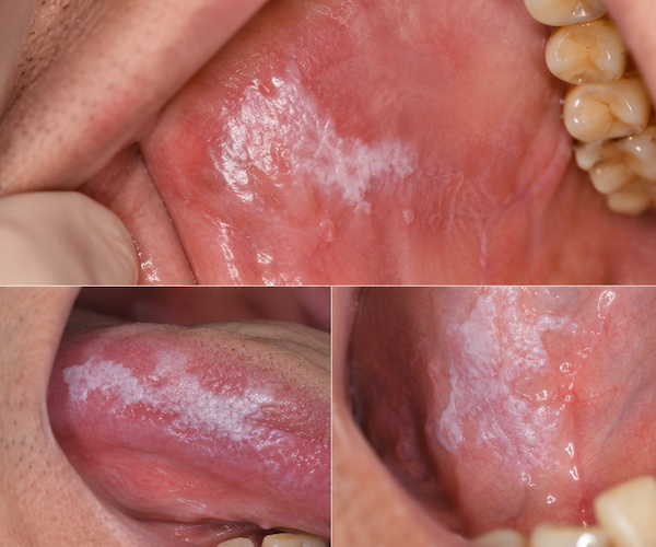
In simple terms, oral leukoplakia refers to a white patch or plaque that:
- Cannot be rubbed off
- Cannot be identified as another oral disease
- Persists over time
- Requires professional evaluation
Therefore, the goal is not fear. Instead, the goal is awareness, early detection, and appropriate management.
Understanding the condition can help patients make informed decisions while avoiding unnecessary panic.
What Is Oral Leukoplakia?
Oral leukoplakia is a clinical diagnosis rather than a single disease.
Dentists use this term when they observe a persistent white lesion on the oral mucosa that cannot be explained by another known condition.
These lesions may appear on:
- The tongue
- The floor of the mouth
- The cheeks
- The gums
- The soft palate
- The corners of the mouth
The appearance varies from patient to patient.
Some lesions remain stable for years. Others gradually change in size or texture.
Importantly, oral leukoplakia itself does not always cause pain.
Many patients only discover it during:
- Routine dental examinations
- Professional cleanings
- Orthodontic evaluations
- Self-examination in a mirror
Because symptoms are often minimal, regular dental checkups remain extremely important.
Two Types of Oral Leukoplakia
Not all leukoplakia lesions carry the same level of risk.
Clinically, dentists often divide oral leukoplakia into two major categories.
Homogeneous Leukoplakia
This is the more common form.
Characteristics include:
- Flat surface
- Uniform white appearance
- Clear boundaries
- Smooth or slightly wrinkled texture
Generally, homogeneous lesions have a lower risk of malignant transformation.
Non-Homogeneous Leukoplakia
This type requires closer attention.
Characteristics include:
- Rough surface
- Raised areas
- Granular appearance
- Ulceration
- Verrucous or wart-like growth
Research consistently shows that non-homogeneous leukoplakia carries a significantly higher cancer risk.
Therefore, early assessment becomes even more important when these features are present.
Which White Patches Have a Higher Cancer Risk?
One of the most common questions patients ask is whether oral leukoplakia will become cancer.
The answer is not straightforward.
Studies report malignant transformation rates ranging from approximately 0.13% to 17.5% during long-term follow-up.
Although the overall risk varies, several factors increase concern.
High-Risk Locations
Certain areas of the mouth have a higher likelihood of malignant transformation.
These include:
- Lateral border of the tongue
- Underside of the tongue
- Floor of the mouth
- Soft palate
- Inner corner of the mouth
These tissues experience rapid cellular turnover and may be more vulnerable to abnormal changes.
Non-Homogeneous Lesions
As discussed earlier, irregular lesions have a higher cancer risk than smooth white plaques.
Candida or HPV Infection
The presence of fungal infection or HPV may increase irritation and accelerate disease progression.
Large Lesion Size
Lesions larger than 200 mm² deserve closer monitoring.
Generally, larger lesions have a higher risk profile.
Long Disease Duration
The longer a lesion remains untreated, the greater the concern for progression.
Female Patients Without Traditional Risk Factors
Interestingly, some studies suggest that leukoplakia occurring in non-smokers and female patients may carry a relatively higher malignant potential.
Epithelial Dysplasia
Biopsy findings play the most important role in risk assessment.
Pathologists classify dysplasia as:
- Mild
- Moderate
- Severe
Severe epithelial dysplasia is considered a true precancerous condition and requires prompt intervention.
How Is Oral Leukoplakia Diagnosed?
Diagnosis begins with a thorough clinical examination.
However, appearance alone cannot determine cancer risk.
Therefore, dentists often use several diagnostic methods.
Clinical Evaluation
The dentist assesses:
- Size
- Color
- Surface texture
- Location
- Duration
Medical History Review
Risk factors are reviewed, including:
- Smoking
- Alcohol use
- Betel nut chewing
- Previous oral lesions
- Family history
Biopsy Examination
A biopsy remains the gold standard.
A small tissue sample is examined under a microscope.
This allows specialists to determine:
- Presence of dysplasia
- Degree of abnormality
- Need for treatment
At Huangshan International Dental Hospital, advanced AI-assisted diagnostic systems help improve imaging analysis and treatment planning. This technology allows clinicians to identify suspicious changes more accurately and efficiently.
Treatment Focus: Reduce Risk and Improve Comfort
At present, there is no guaranteed cure for oral leukoplakia.
Therefore, treatment focuses on controlling progression and reducing cancer risk.
Eliminate Irritating Factors
The first step is removing ongoing irritation.
Patients should:
- Stop smoking completely
- Avoid alcohol consumption
- Stop chewing betel nut
- Improve oral hygiene
- Repair broken teeth
- Replace poorly fitting dentures
Reducing chronic irritation can significantly improve outcomes.
Medication Therapy
Some patients may benefit from:
- Topical retinoids
- Vitamin A derivatives
- Beta-carotene supplements
- Vitamin E
- Antioxidant therapy
However, treatment should always be supervised by a dental professional.
Surgical Removal
For high-risk lesions, surgery may be recommended.
Common indications include:
- Non-homogeneous lesions
- High-risk locations
- Severe dysplasia
- Rapid lesion growth
Even after removal, continued monitoring remains essential because recurrence can occur.
Physical Therapies
Several minimally invasive approaches are available.
These include:
- Cryotherapy
- Laser therapy
- Photodynamic therapy (PDT)
Photodynamic therapy has become increasingly popular because it:
- Preserves healthy tissue
- Produces minimal scarring
- Requires little recovery time
- Can be repeated when necessary
Why Long-Term Follow-Up Is Essential
Many patients mistakenly believe treatment ends once the lesion disappears.
Unfortunately, that is not always true.
Even successfully treated leukoplakia can recur.
Therefore, lifelong monitoring is often recommended.
Regular follow-up appointments allow dentists to:
- Detect recurrence early
- Monitor suspicious changes
- Perform biopsies when needed
- Adjust treatment plans
At Huangshan International Dental Hospital, patients receive personalized follow-up schedules based on individual risk factors. Furthermore, the hospital’s multidisciplinary dental team collaborates across oral medicine, pathology, periodontics, and restorative dentistry to provide comprehensive care for complex cases.
Frequently Asked Questions
Is oral leukoplakia always cancer?
No. Most cases are not cancer. However, some lesions can develop into oral cancer over time.
Can oral leukoplakia disappear on its own?
Sometimes lesions improve after removing irritants such as tobacco or poorly fitting dentures. However, professional monitoring remains necessary.
What does oral leukoplakia look like?
It typically appears as a white patch or plaque that cannot be rubbed off and persists over time.
Is oral leukoplakia painful?
Many lesions are painless. Some patients experience irritation, burning, or sensitivity.
Should every leukoplakia lesion be biopsied?
Not necessarily. However, suspicious or persistent lesions often require biopsy to assess cancer risk.
Conclusion
Oral leukoplakia is one of the most common potentially malignant oral disorders, but finding a white patch in your mouth does not automatically mean cancer.
The key is early evaluation, accurate diagnosis, and consistent follow-up. While some lesions remain stable for years, others carry a higher risk of malignant transformation. Factors such as lesion type, location, size, infection status, and biopsy findings all influence prognosis.
If you notice a persistent white patch that does not rub off, do not ignore it. At the same time, do not assume the worst. Schedule a professional examination and follow your dentist’s recommendations. Early action remains the most effective strategy for protecting long-term oral health.