
Bitewing x-rays are one of the maximum not unusualplace dental radiographs, gambling a important position withinside the detection of enamel decay and the evaluation of dental restorations in recurring dental care. With their cappotential to focus on precise regions of the mouth, particularly the returned enamel, bitewing x-rays are a cornerstone for preventive dentistry. However, for plenty sufferers or even the ones new to dental care, a key query arises: which enamel have to truly be seen on a bitewing x-ray, and why is this option so critical for correct diagnosis?
Understanding Bitewing Radiography
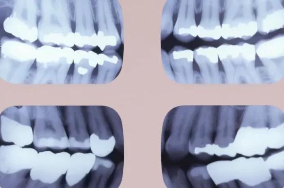
Bitewing radiography is a way used to seize snap shots of the crowns of your posterior enamel, specially your premolars and molars. The name “bitewing” comes from the unique method wherein sufferers bit down on a small wing or tab connected to the movie withinside the mouth. Modern bitewing strategies on the whole use holders with plastic systems and virtual sensors, making the technique extra snug and snap shots clearer.
This x-ray is specially designed to recognition at the regions wherein cavities are maximum probable to develop: among the returned enamel. The bitewing is specific amongst dental x-rays as it isn’t supposed to expose the enamel roots or the place close to the top of the roots, however instead it is supposed to offer an in depth go phase of the top and decrease enamel wherein they contact every different.
Which Teeth Should a Bitewing X-Ray Include?
Focus on Premolars and Molars
A conventional bitewing x-ray targets to in reality display the crowns of each top and decrease premolars and molars. The fundamental intention is to seize the regions wherein those returned enamel contact, due to the fact those touch factors are enormously vulnerable to decay and are regularly invisible at some point of a preferred visible exam.
In maximum dental practices, a hard and fast of 4 bitewing x-rays is taken— on every facet of the mouth. The standard insurance includes:
- Premolars (additionally called bicuspids): First and 2nd premolars are a first-rate recognition due to the fact those are transitional enamel among the dog (the “eye enamel”) and primary molar.
- Molars: First and 2nd molars are crucial for checking decay and current restorations like fillings and crowns. If a 3rd molar (expertise enamel) is gift, its inclusion relies upon on its alignment and relevance to the patient’s oral health.
- The bitewing have to normally begin simply distal (behind) to the dog enamel, preferably catching the distal fringe of the dog as a landmark. The photo then extends all of the manner to the returned of the arch, overlaying to the final gift molar, that’s regularly the second one or 1/3 molar for the patient.
Front enamel (incisors and canines) aren’t proven on bitewing x-rays due to the fact they’re much less susceptible to the sorts of problems bitewings are designed to locate and require different sorts of x-rays, along with periapical or anterior films.
Why Are These Teeth Selected?
The choice of enamel is primarily based totally on medical need. Bitewing radiographs are tasked more often than not with locating interproximal decay (cavities among enamel), assessing how current restorations are carrying or leaking, and checking for early symptoms and symptoms of bone loss associated with periodontal disease. The touch regions of the returned enamel are the maximum hard to peer visually, making x-ray exam essential.
If the bitewing does now no longer cowl all of the molars or misses the premolars, crucial regions may work unseen, probably main to ignored diagnoses of cavities or problems beneathneath current crowns and fillings.
Separate Images for Complete Coverage
Because the dental arches curve, it’s far regularly not possible to get each the premolars and molars in sharp parallel recognition on a unmarried picture. Thus, the same old protocol is to take photographs in step with side: the first (premolar bitewing) is located to seize from the distal of the dog to the second one premolar, whilst the second one (molar bitewing) is located to picture the first, second, and probably 1/3 molars.
Technical Requirements for Accurate Bitewing Images
Patient and Sensor Positioning
For accurate results, the picture receptor (virtual sensor or film) have to be located parallel to the enamel in question. The top and decrease edges of the sensor have to be in step with the biting surfaces, and the sensor have to be near the enamel to restriction distortion. The x-ray beam have to be aimed immediately via the touch factors of the enamel at a horizontal and vertical perspective that forestalls overlapping the photographs of adjoining enamel.
Common Challenges in Image Acquisition
Several limitations can save you right imaging. Variations in dental arch length—specifically in youngsters or people with small jaws—might also additionally restriction picture clarity. Sometimes, holders are adjusted or the “tab” technique is used, in which the affected person bites on a sticking out tab in preference to a inflexible platform. In uncommon instances, anatomical variations suggest the whole premolar or molar vicinity might not suit in a single view inspite of separate photographs, wherein case extra perspectives can be necessary.
Bitewing X-Rays in Special Circumstances
Children and Mixed Dentition
For youngsters or at some stage in the blended dentition phase, bitewing radiographs are adjusted to consist of the particular returned enamel present. Only enamel in purposeful touch (opposing top and decrease enamel that meet at some stage in biting) are imaged, as those are the zones maximum prone to caries. For very younger youngsters with best primary (baby) molars, a smaller sensor length is used to offer the first-class suit and comfort.
Patients With Missing Teeth or Crowding
If a affected person is lacking a few molars or premolars, the bitewing remains located to pattern as many posterior contacts as possible. In instances with crowding or overlapping, bitewings are cautiously angled to maximise separation of touch factors, enhancing detection of hidden cavities.
The Role of Bitewing X-Rays in Diagnosis
Bitewing radiographs are taken into consideration important for:
- Detecting new cavities among returned tooth early, earlier than they reason symptoms
- Checking for gaps or leaks below vintage fillings and crowns
- Monitoring bone stage among tooth, offering early signs of gum disease
- Helping dentists music the development of dental caries and the steadiness of present dental paintings over time
These photographs are commonly now no longer appropriate for comparing enamel roots or the helping bone on the tip of the root. For those issues, periapical x-rays or panoramic movies are used.
How Often Should Bitewings Be Taken?
Routine bitewing x-rays are usually endorsed as soon as a 12 months for adults and kids at regular threat for decay. For the ones at better threat, such as though a affected person has a couple of current cavities or great dental paintings, screening can be endorsed each six months. Your dentist will make the very last advice primarily based totally in your fame and dental history.
Conclusion
The accurate tooth to be covered on a bitewing x-ray are the top and decrease premolars and molars on every aspect of the mouth. This standardized insurance guarantees that the touch factors maximum at threat for decay are visualized effectively, helping correct and well timed diagnosis. Consistent technique, right sensor placement, and attention for affected person-unique anatomy are important for high-quality, diagnostic photographs.
Frequently Asked Questions (FAQs)
Which tooth are covered in a preferred bitewing x-ray?
A preferred bitewing indicates the crowns of the top and decrease premolars and molars, beginning simply in the back of the dog enamel and increasing to the remaining gift molar.
Why are premolars and molars the point of interest of bitewings and now no longer the front tooth?
Premolars and molars have aspect surfaces (interproximal areas) in which cavities most customarily develop, and those are tough to take a look at visually. Front tooth are much less liable to those troubles and are imaged with different sorts of x-rays.
How frequently do I want bitewing x-rays?
Most sufferers want bitewing x-rays as soon as a 12 months, however your dentist can also additionally advocate them extra or much less frequently primarily based totally in your oral fitness and history.
Can bitewing x-rays locate gum disease?
Bitewings can display modifications in bone stage among the tooth, which facilitates withinside the early detection of gum disease, however aren’t complete for root or complete bone assessment.
Are bitewing x-rays safe?
Yes, current bitewing x-rays use virtual sensors that require very low degrees of radiation, and dental experts take precautions to preserve publicity to a minimal for affected person safety.